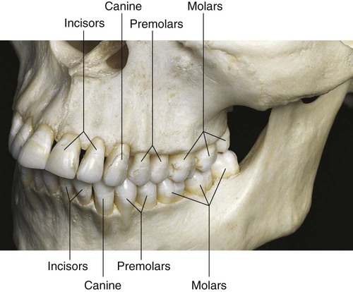
1 Clinical Significance Of Dental Anatomy Histology Physiology And

Find inspiration for 1 Clinical Significance Of Dental Anatomy Histology Physiology And with our image finder website, 1 Clinical Significance Of Dental Anatomy Histology Physiology And is one of the most popular images and photo galleries in Histology Of Incisal And Molar Regions In The Wildtype And Gallery, 1 Clinical Significance Of Dental Anatomy Histology Physiology And Picture are available in collection of high-quality images and discover endless ideas for your living spaces, You will be able to watch high quality photo galleries 1 Clinical Significance Of Dental Anatomy Histology Physiology And.
aiartphotoz.com is free images/photos finder and fully automatic search engine, No Images files are hosted on our server, All links and images displayed on our site are automatically indexed by our crawlers, We only help to make it easier for visitors to find a free wallpaper, background Photos, Design Collection, Home Decor and Interior Design photos in some search engines. aiartphotoz.com is not responsible for third party website content. If this picture is your intelectual property (copyright infringement) or child pornography / immature images, please send email to aiophotoz[at]gmail.com for abuse. We will follow up your report/abuse within 24 hours.
Related Images of 1 Clinical Significance Of Dental Anatomy Histology Physiology And
Histology Of Incisal And Molar Regions In The Wildtype And
Histology Of Incisal And Molar Regions In The Wildtype And
850×1324
Morphology Of Adult Wildtype And Vimentin Deficient Wounds A Wax
Morphology Of Adult Wildtype And Vimentin Deficient Wounds A Wax
795×1048
Fgf Genetic Interactions In Incisor And Molar Development Ai Lower
Fgf Genetic Interactions In Incisor And Molar Development Ai Lower
671×949
Figure S8 Histology Of D5 Maxillary Molars Hande Stained Sections 10×
Figure S8 Histology Of D5 Maxillary Molars Hande Stained Sections 10×
850×1042
Sagittal Sections Of Mandibular Incisors Of Wild Type Wt And
Sagittal Sections Of Mandibular Incisors Of Wild Type Wt And
489×505
Figure S9 Histology Of D11 Maxillary Molars Hande Stained Sections
Figure S9 Histology Of D11 Maxillary Molars Hande Stained Sections
850×1035
Pone 0055274 G006scube3 Is Expressed In Multiple Tissues During
Pone 0055274 G006scube3 Is Expressed In Multiple Tissues During
512×536
Immunolocalization For Type I Collagen And Fibronectin In The Wild Type
Immunolocalization For Type I Collagen And Fibronectin In The Wild Type
850×695
Morphology Of The Dentition Of Amelx Mutant Mice In Wild Type Wt
Morphology Of The Dentition Of Amelx Mutant Mice In Wild Type Wt
560×534
Maxillary D5 First Molar Histology A Low Magnification Sections Of
Maxillary D5 First Molar Histology A Low Magnification Sections Of
850×1113
Skin Histopathology Overview A The Dorsal Skin Of 44 Wild Type Mice
Skin Histopathology Overview A The Dorsal Skin Of 44 Wild Type Mice
850×820
Ppt Tooth Histology And Morphology Powerpoint Presentation Free
Ppt Tooth Histology And Morphology Powerpoint Presentation Free
1024×768
1 Oral Structures And Tissues Pocket Dentistry
1 Oral Structures And Tissues Pocket Dentistry
1375×783
Histology 7 Week Mmp20−− Mandibular Incisor A Basal Ends Of
Histology 7 Week Mmp20−− Mandibular Incisor A Basal Ends Of
850×953
Appearance Of Odaph Odaphc41 And Odaphc41c41 Incisors And
Appearance Of Odaph Odaphc41 And Odaphc41c41 Incisors And
850×1015
Pdf Altered Distribution Of Extracellular Matrix Proteins In The
Pdf Altered Distribution Of Extracellular Matrix Proteins In The
640×640
Hande Staining Of The Mandibular First Molars From 1 Day Old P1
Hande Staining Of The Mandibular First Molars From 1 Day Old P1
850×482
Histology Of Retinas Of 30 Week Old Heterozygous And Wildtype Mice Low
Histology Of Retinas Of 30 Week Old Heterozygous And Wildtype Mice Low
850×1248
Figure S11b Histology Of 7 Week Wdr72 Mandibular Incisor 9 A
Figure S11b Histology Of 7 Week Wdr72 Mandibular Incisor 9 A
850×992
Alterations Of Enamel In Molars And The Molar Root Phenotype In Latent
Alterations Of Enamel In Molars And The Molar Root Phenotype In Latent
850×1258
Histochemistry And Far Western Immunohistochemistry With A −
Histochemistry And Far Western Immunohistochemistry With A −
850×1101
An Increasing Gradient Of Ki 67 Staining In The Incisal To Apical
An Increasing Gradient Of Ki 67 Staining In The Incisal To Apical
748×553
1 Clinical Significance Of Dental Anatomy Histology Physiology And
1 Clinical Significance Of Dental Anatomy Histology Physiology And
500×414
Three Dimensional Micro Ct Images Of The Upper And Lower First Molar
Three Dimensional Micro Ct Images Of The Upper And Lower First Molar
850×853
Histological Characteristics Of The Right Maxillary First Molar 16
Histological Characteristics Of The Right Maxillary First Molar 16
850×617
Histopathology Of The Left Mandibular Primary Second Molar Decalcified
Histopathology Of The Left Mandibular Primary Second Molar Decalcified
508×320
Wild Type Control Tg Mmp20 Incisors Had A Sharp Incisal Tip And
Wild Type Control Tg Mmp20 Incisors Had A Sharp Incisal Tip And
722×1110
Ppt Tooth Histology And Morphology Powerpoint Presentation Free
Ppt Tooth Histology And Morphology Powerpoint Presentation Free
1024×768
