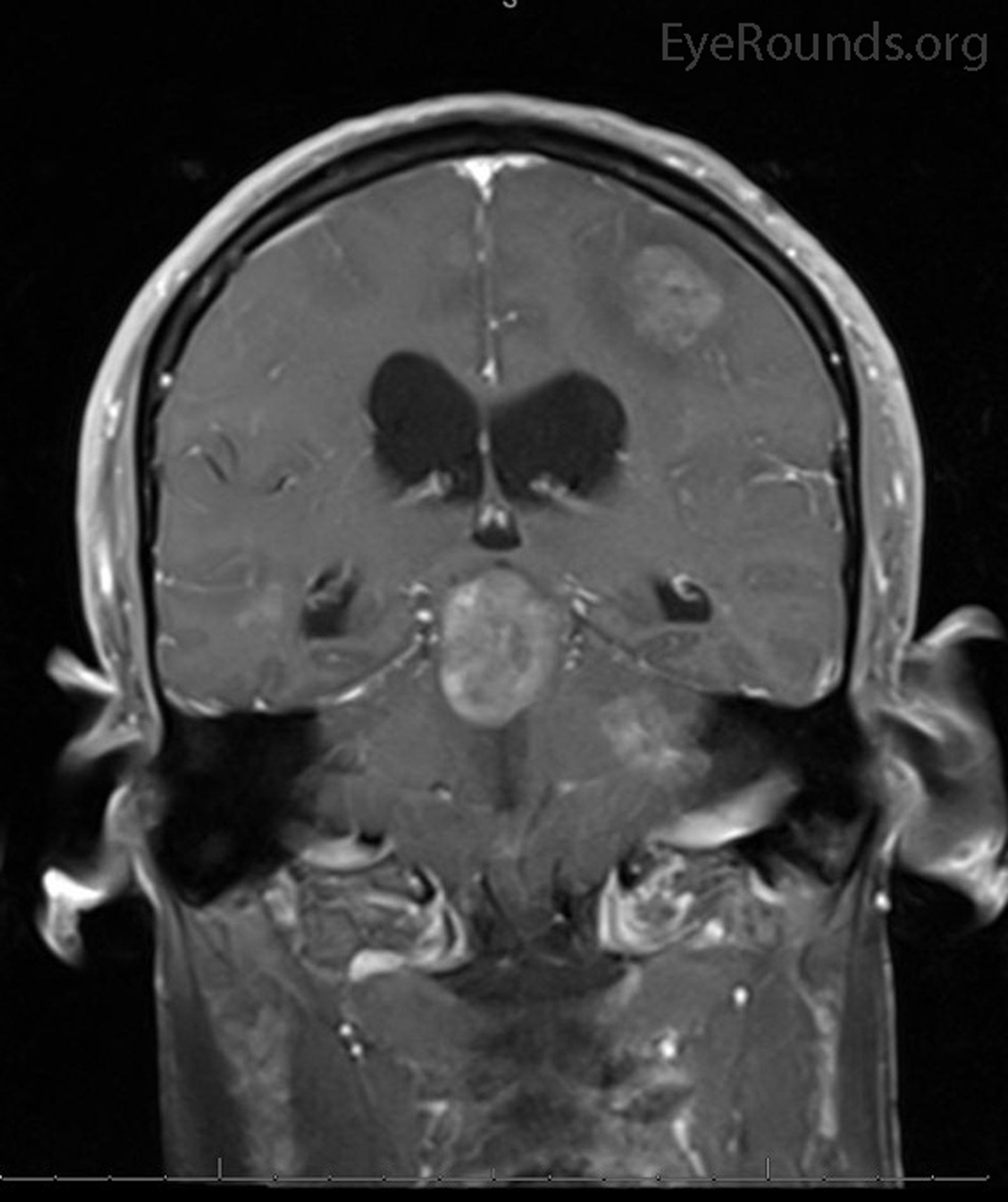
Bilateral Internuclear Ophthalmoplegia And Thalamic Esotropia The

Find inspiration for Bilateral Internuclear Ophthalmoplegia And Thalamic Esotropia The with our image finder website, Bilateral Internuclear Ophthalmoplegia And Thalamic Esotropia The is one of the most popular images and photo galleries in Coronal Mri With Contrast Done After Radiosurgery To The Suprasellar Gallery, Bilateral Internuclear Ophthalmoplegia And Thalamic Esotropia The Picture are available in collection of high-quality images and discover endless ideas for your living spaces, You will be able to watch high quality photo galleries Bilateral Internuclear Ophthalmoplegia And Thalamic Esotropia The.
aiartphotoz.com is free images/photos finder and fully automatic search engine, No Images files are hosted on our server, All links and images displayed on our site are automatically indexed by our crawlers, We only help to make it easier for visitors to find a free wallpaper, background Photos, Design Collection, Home Decor and Interior Design photos in some search engines. aiartphotoz.com is not responsible for third party website content. If this picture is your intelectual property (copyright infringement) or child pornography / immature images, please send email to aiophotoz[at]gmail.com for abuse. We will follow up your report/abuse within 24 hours.
Related Images of Bilateral Internuclear Ophthalmoplegia And Thalamic Esotropia The
Mri In Ms Stroke Brain Tumor Fieldstrength Mri Philips Healthcare
Mri In Ms Stroke Brain Tumor Fieldstrength Mri Philips Healthcare
700×400
T1 Coronal Mri With Contrast Highlighting The Cavernous Sinus
T1 Coronal Mri With Contrast Highlighting The Cavernous Sinus
475×550
Coronal T2 Weighted Magnetic Resonance Image Of The Brain The Bmj
Coronal T2 Weighted Magnetic Resonance Image Of The Brain The Bmj
1152×1280
Figure 16 A Coronal T2 Weighted Image Endotext Ncbi Bookshelf
Figure 16 A Coronal T2 Weighted Image Endotext Ncbi Bookshelf
766×959
Figure 28 T1 Weighted Coronal Enhanced Image Endotext Ncbi
Figure 28 T1 Weighted Coronal Enhanced Image Endotext Ncbi
676×473
Coronal Magnetic Resonance Imaging Mri Of The Pelvis Open I
Coronal Magnetic Resonance Imaging Mri Of The Pelvis Open I
512×259
Orbital Eosinophilic Granuloma Ophthalmology The
Orbital Eosinophilic Granuloma Ophthalmology The
1136×1363
Jcm Free Full Text Evaluation Of The Value Of Perfusion Weighted
Jcm Free Full Text Evaluation Of The Value Of Perfusion Weighted
2383×2327
Cavernous Sinus Syndrome Secondary To Pituitary Apoplexy
Cavernous Sinus Syndrome Secondary To Pituitary Apoplexy
2000×1592
Role For Stereotactic Radiosurgery In Pituitary Dependent Cushing
Role For Stereotactic Radiosurgery In Pituitary Dependent Cushing
632×319
Atypical Pituitary Adenomas 10 Years Of Experience In A Reference
Atypical Pituitary Adenomas 10 Years Of Experience In A Reference
1024×617
Bilateral Internuclear Ophthalmoplegia And Thalamic Esotropia The
Bilateral Internuclear Ophthalmoplegia And Thalamic Esotropia The
2000×2383
T1 Weighted Coronal Mri Scan Of The Pelvis Post Partum Open I
T1 Weighted Coronal Mri Scan Of The Pelvis Post Partum Open I
512×512
Shoulder Mri Coronal 1 Anterior Diagram Quizlet
Shoulder Mri Coronal 1 Anterior Diagram Quizlet
512×512
Dxi 2 10 Pelvis And Hip Coronal Mri 3 Diagram Quizlet
Dxi 2 10 Pelvis And Hip Coronal Mri 3 Diagram Quizlet
1024×757
Role For Stereotactic Radiosurgery In Pituitary Dependent Cushing
Role For Stereotactic Radiosurgery In Pituitary Dependent Cushing
632×358
Diagnostics Free Full Text Ischiofemoral Impingement Syndrome
Diagnostics Free Full Text Ischiofemoral Impingement Syndrome
3806×1301
Coronal Contrast Enhanced T1 Weighted Brain Mri An Enh Open I
Coronal Contrast Enhanced T1 Weighted Brain Mri An Enh Open I
512×630
Stereotactic Radiosurgery In The Management Of Acoustic Neuromas
Stereotactic Radiosurgery In The Management Of Acoustic Neuromas
1024×772
Diagram Of Coronal Brain Mri 4th Ventricle Quizlet
Diagram Of Coronal Brain Mri 4th Ventricle Quizlet
587×610
Mri Brain Coronal Cross Sectional Anatomy Image Brain Anatomy Mri
Mri Brain Coronal Cross Sectional Anatomy Image Brain Anatomy Mri
800×550
Normal Coronal Mri Of The Brain Photograph By Medical Body Scans
Normal Coronal Mri Of The Brain Photograph By Medical Body Scans
900×900
Coronal T1 Mri Lateral And 4th Ventricle Diagram Quizlet
Coronal T1 Mri Lateral And 4th Ventricle Diagram Quizlet
508×640
Endolymphatic Sac Tumor Tanner P Lyons Laura Barry Robert T
Endolymphatic Sac Tumor Tanner P Lyons Laura Barry Robert T
1774×1454
T2 Flair Coronal Mri Through The Pons And 3rd Ventricle
T2 Flair Coronal Mri Through The Pons And 3rd Ventricle
475×550
Tomography Free Full Text T1 Weighted Contrast Enhancement
Tomography Free Full Text T1 Weighted Contrast Enhancement
3651×2443
Orbital Eosinophilic Granuloma Ophthalmology The
Orbital Eosinophilic Granuloma Ophthalmology The
1200×1439
Radiosurgery For Chordomas And Chondrosarcomas Of The Skull Base In
Radiosurgery For Chordomas And Chondrosarcomas Of The Skull Base In
501×286
