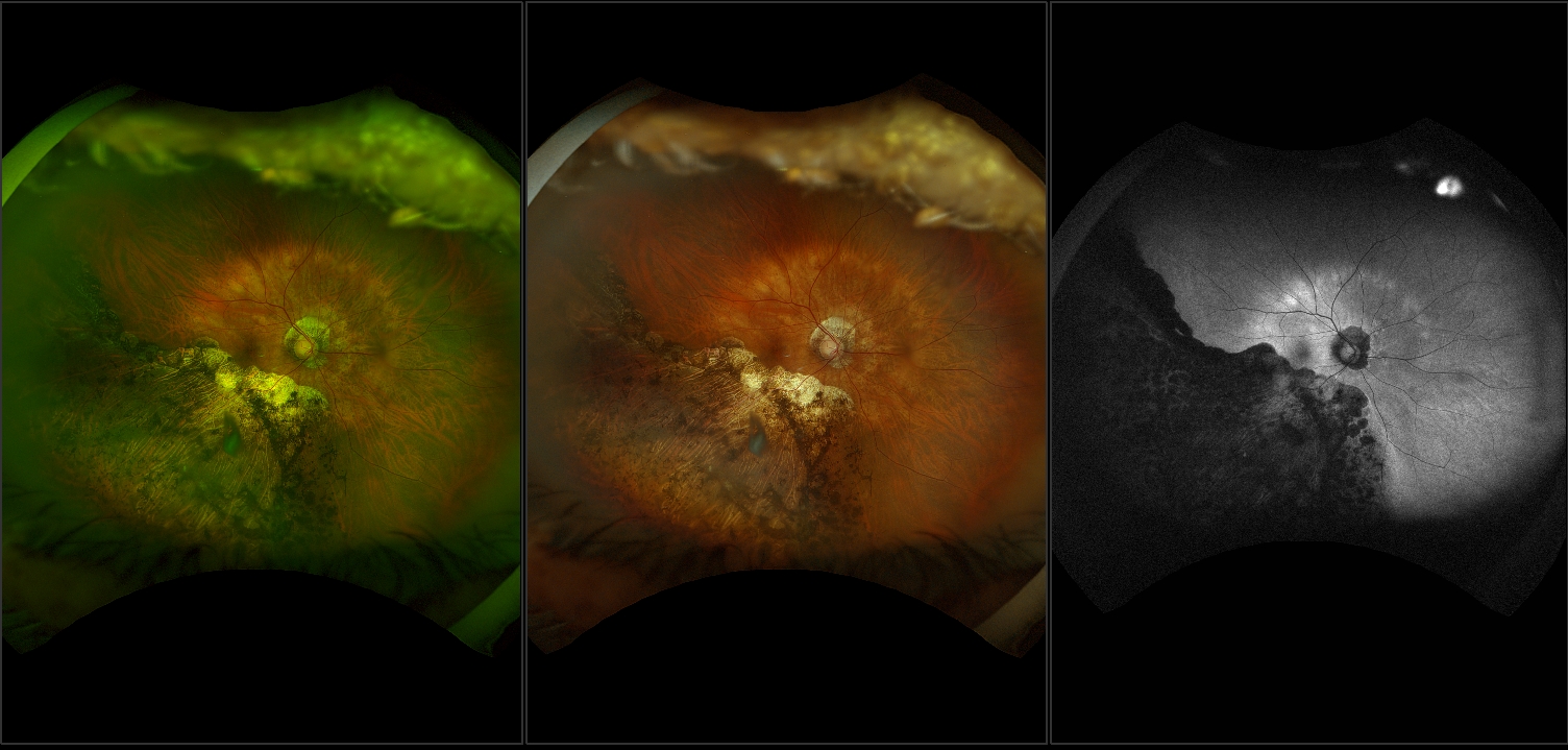
California Peripapillary Atrophy With Chronic Partial Retinal

Find inspiration for California Peripapillary Atrophy With Chronic Partial Retinal with our image finder website, California Peripapillary Atrophy With Chronic Partial Retinal is one of the most popular images and photo galleries in California Peripapillary Atrophy With Chronic Partial Retinal Gallery, California Peripapillary Atrophy With Chronic Partial Retinal Picture are available in collection of high-quality images and discover endless ideas for your living spaces, You will be able to watch high quality photo galleries California Peripapillary Atrophy With Chronic Partial Retinal.
aiartphotoz.com is free images/photos finder and fully automatic search engine, No Images files are hosted on our server, All links and images displayed on our site are automatically indexed by our crawlers, We only help to make it easier for visitors to find a free wallpaper, background Photos, Design Collection, Home Decor and Interior Design photos in some search engines. aiartphotoz.com is not responsible for third party website content. If this picture is your intelectual property (copyright infringement) or child pornography / immature images, please send email to aiophotoz[at]gmail.com for abuse. We will follow up your report/abuse within 24 hours.
Related Images of California Peripapillary Atrophy With Chronic Partial Retinal
California Peripapillary Atrophy With Chronic Partial Retinal
California Peripapillary Atrophy With Chronic Partial Retinal
1504 x 719 · JPG
Optic Nerve Evaluation In Glaucoma California Optometric Association
Optic Nerve Evaluation In Glaucoma California Optometric Association
1500 x 1105 · JPG
Peripapillary Atrophy American Academy Of Ophthalmology
Peripapillary Atrophy American Academy Of Ophthalmology
975 x 700 ·
Peripapillary Chorioretinal Atrophy Semantic Scholar
Peripapillary Chorioretinal Atrophy Semantic Scholar
646 x 490 · png
A Color Fundus Photograph Of The Right Eye Reveals Peripapillary
A Color Fundus Photograph Of The Right Eye Reveals Peripapillary
702 x 750 · png
Figure 1 From Peripapillary Retinal Nerve Fiber Layer Swelling Predicts
Figure 1 From Peripapillary Retinal Nerve Fiber Layer Swelling Predicts
1380 x 422 · png
A Reactive Peripapillary Atrophy And Pigmentary Alteration In The
A Reactive Peripapillary Atrophy And Pigmentary Alteration In The
850 x 1471 · JPG
Case 2 Fundus Photographs A And B Reveal Peripapillary Atrophy In Both
Case 2 Fundus Photographs A And B Reveal Peripapillary Atrophy In Both
850 x 736 · JPG
Figure 1 From Peripapillary Atrophy Detection By Sparse Biologically
Figure 1 From Peripapillary Atrophy Detection By Sparse Biologically
1142 x 518 · png
Ultra Wide Field Fundus Image A And Blue Light Autofluorescence C
Ultra Wide Field Fundus Image A And Blue Light Autofluorescence C
850 x 933 · JPG
Symptomatic Branch Retinal Artery Occlusion An Under Recognized Sign
Symptomatic Branch Retinal Artery Occlusion An Under Recognized Sign
2000 x 1125 · JPG
Associations Of Peripapillary Atrophy And Fundus Tessellation With
Associations Of Peripapillary Atrophy And Fundus Tessellation With
3445 x 1158 · JPG
Figure 2 From Peripapillary Retinal Nerve Fiber Layer Swelling Predicts
Figure 2 From Peripapillary Retinal Nerve Fiber Layer Swelling Predicts
898 x 696 · png
California Peripapillary Pachychoroid Syndrome Rg Rgb Af Icg
California Peripapillary Pachychoroid Syndrome Rg Rgb Af Icg
2500 x 1132 · JPG
Case 1 A Color Fundus Image Of The Right Eye With Peripapillary
Case 1 A Color Fundus Image Of The Right Eye With Peripapillary
789 x 566 · JPG
Peripapillary Choroidal Cavitation As A Feature Of Pathological Myopia
Peripapillary Choroidal Cavitation As A Feature Of Pathological Myopia
1280 x 1037 · JPG
Peripapillary Scarring Associated With Atrophic Macular Lesion
Peripapillary Scarring Associated With Atrophic Macular Lesion
643 x 416 · JPG
Peripapillary And Macular Retinoschisis Associated With Advanced
Peripapillary And Macular Retinoschisis Associated With Advanced
1660 x 2489 · JPG
A Fundus Photography Od Of Case 2 Showing A Peripapillary
A Fundus Photography Od Of Case 2 Showing A Peripapillary
850 x 567 · png
Peripapillary Staphyloma Retina Image Bank
Peripapillary Staphyloma Retina Image Bank
643 x 540 · JPG
Peripapillary Atrophy With High Myopia Retina Image Bank
Peripapillary Atrophy With High Myopia Retina Image Bank
643 x 428 · JPG
Exudation In Peripapillary Retina Due To Syphilis Retina Image Bank
Exudation In Peripapillary Retina Due To Syphilis Retina Image Bank
643 x 643 · JPG
Atlas Entry Peripapillary Combined Hamartoma Of The Retina And
Atlas Entry Peripapillary Combined Hamartoma Of The Retina And
686 x 720 · JPG
Peripapillary Atrophy In Primary Angle Closure Glaucoma A Comparative
Peripapillary Atrophy In Primary Angle Closure Glaucoma A Comparative
3760 x 2171 · JPG
Images Of β Peripapillary Atrophy In A Fundus Colour Paragraph A D
Images Of β Peripapillary Atrophy In A Fundus Colour Paragraph A D
600 x 464 · JPG
Microstructure Of Peripapillary Atrophy And Subsequent Visual Field
Microstructure Of Peripapillary Atrophy And Subsequent Visual Field
590 x 652 · JPG
