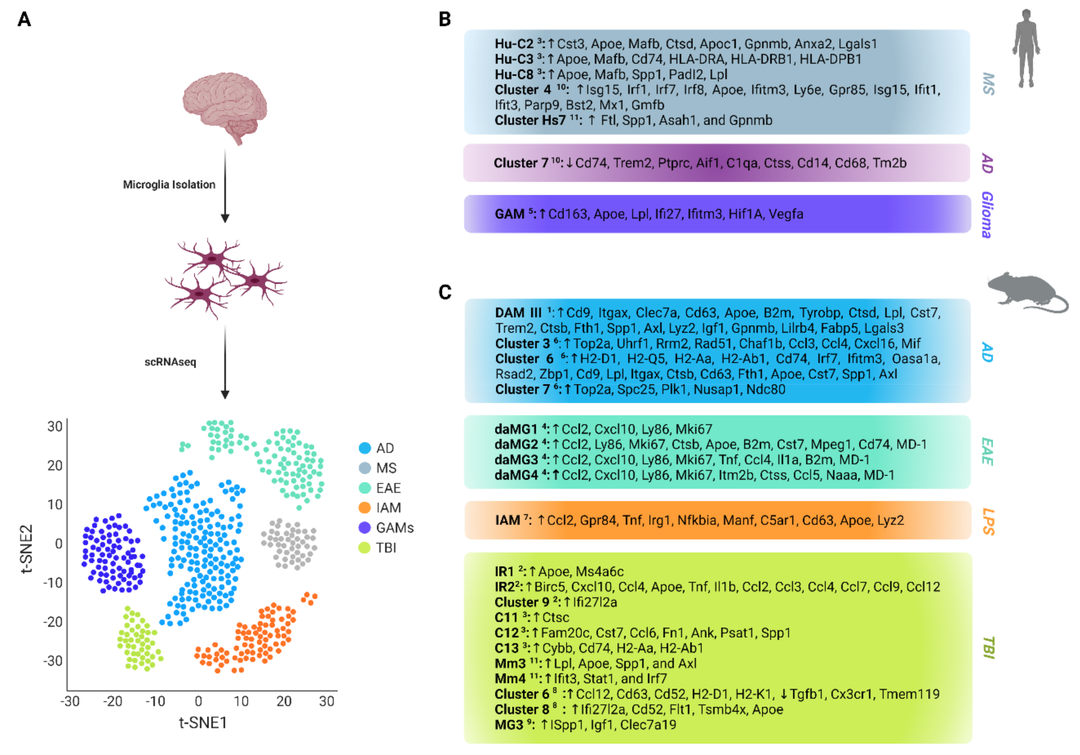
Cells Free Full Text Profiling Microglia Through Single Cell Rna

Find inspiration for Cells Free Full Text Profiling Microglia Through Single Cell Rna with our image finder website, Cells Free Full Text Profiling Microglia Through Single Cell Rna is one of the most popular images and photo galleries in Rat Microglial Cells Express T Cell Markers Microglial Cells Iba1 Gallery, Cells Free Full Text Profiling Microglia Through Single Cell Rna Picture are available in collection of high-quality images and discover endless ideas for your living spaces, You will be able to watch high quality photo galleries Cells Free Full Text Profiling Microglia Through Single Cell Rna.
aiartphotoz.com is free images/photos finder and fully automatic search engine, No Images files are hosted on our server, All links and images displayed on our site are automatically indexed by our crawlers, We only help to make it easier for visitors to find a free wallpaper, background Photos, Design Collection, Home Decor and Interior Design photos in some search engines. aiartphotoz.com is not responsible for third party website content. If this picture is your intelectual property (copyright infringement) or child pornography / immature images, please send email to aiophotoz[at]gmail.com for abuse. We will follow up your report/abuse within 24 hours.
Related Images of Cells Free Full Text Profiling Microglia Through Single Cell Rna
Rat Microglial Cells Express T Cell Markers Microglial Cells Iba1
Rat Microglial Cells Express T Cell Markers Microglial Cells Iba1
603×402
Iba1 Microglia Colonize The Rat Proliferative Zones A At E13 Very Few
Iba1 Microglia Colonize The Rat Proliferative Zones A At E13 Very Few
850×1165
Iba1 Immunoreative Microglial Cells In The Spinal Cords Of Sci Rats At
Iba1 Immunoreative Microglial Cells In The Spinal Cords Of Sci Rats At
850×861
The Microglia Marker Iba1 Is Enriched In Podonuts And Podosomes All
The Microglia Marker Iba1 Is Enriched In Podonuts And Podosomes All
600×506
Confocal Images Show Localization Of Proliferating Microglial Cells
Confocal Images Show Localization Of Proliferating Microglial Cells
775×943
The Microglia Marker Iba1 Is Enriched In Podonuts And Podosomes All
The Microglia Marker Iba1 Is Enriched In Podonuts And Podosomes All
640×640
Reduction Of Microgliosis In Hnscs Treated Rats A Representative
Reduction Of Microgliosis In Hnscs Treated Rats A Representative
850×1026
Frontiers Microglia And Astrocyte Function And Communication What Do
Frontiers Microglia And Astrocyte Function And Communication What Do
3161×3677
Frontiers Overview Of General And Discriminating Markers Of
Frontiers Overview Of General And Discriminating Markers Of
1300×946
Frontiers Overview Of General And Discriminating Markers Of
Frontiers Overview Of General And Discriminating Markers Of
1300×1182
Microglial Cells In The Lha Region Of Obese Rats Were Accompanied By
Microglial Cells In The Lha Region Of Obese Rats Were Accompanied By
850×576
Dex Induced Microglial Dysfunction In Rat Primary Microglial Cells The
Dex Induced Microglial Dysfunction In Rat Primary Microglial Cells The
850×1156
Iba1 Microglia Phagocytose Neural Precursor Cells In The Developing
Iba1 Microglia Phagocytose Neural Precursor Cells In The Developing
850×394
Immunofluorescence Staining Using Iba1 And Tmem119 For Microglial
Immunofluorescence Staining Using Iba1 And Tmem119 For Microglial
500×446
Frontiers Microglia Versus Myeloid Cell Nomenclature During Brain
Frontiers Microglia Versus Myeloid Cell Nomenclature During Brain
1000×776
Microglia Regulate The Number Of Neural Precursor Cells In The
Microglia Regulate The Number Of Neural Precursor Cells In The
1158×1280
F480 Iba1 Retinal Microglia And Ciliary Macrophages In The Adult Mouse
F480 Iba1 Retinal Microglia And Ciliary Macrophages In The Adult Mouse
750×1241
Microglial Activation States Markers And Functions Microglia Are
Microglial Activation States Markers And Functions Microglia Are
850×572
In Cultured Rat Microglial Cells The Plasmid Of Human Tau40 Egfp T
In Cultured Rat Microglial Cells The Plasmid Of Human Tau40 Egfp T
640×640
Microglia Have Distinct Expression Signatures Based On Spatial Context
Microglia Have Distinct Expression Signatures Based On Spatial Context
850×1117
Ijms Free Full Text The Role Of Microglia In Modulating
Ijms Free Full Text The Role Of Microglia In Modulating
2972×1924
Frontiers Strategies And Tools For Studying Microglial Mediated
Frontiers Strategies And Tools For Studying Microglial Mediated
1084×725
Effects Of Systemic Injection Of Lps On M1 Microglial Markers
Effects Of Systemic Injection Of Lps On M1 Microglial Markers
850×843
Isolated Microglial Cells Express P2x7 Receptors On Protein And
Isolated Microglial Cells Express P2x7 Receptors On Protein And
640×640
Downregulation Of Stat2 Increased Expression Of Microglial Activation
Downregulation Of Stat2 Increased Expression Of Microglial Activation
850×722
The Number Of Microglial Cells That Express Markers Associated With
The Number Of Microglial Cells That Express Markers Associated With
850×1135
Cells Free Full Text Profiling Microglia Through Single Cell Rna
Cells Free Full Text Profiling Microglia Through Single Cell Rna
3628×2536
A Specific Subset Of Cd163 Cells Does Not Express Classical Microglial
A Specific Subset Of Cd163 Cells Does Not Express Classical Microglial
850×1130
Lps Increases Traf6 And Iba 1 Expression In Primary Microglia Ac
Lps Increases Traf6 And Iba 1 Expression In Primary Microglia Ac
850×739
Rat Cochlear Microglial Cell Line Mocha Kerafast
Rat Cochlear Microglial Cell Line Mocha Kerafast
500×500
