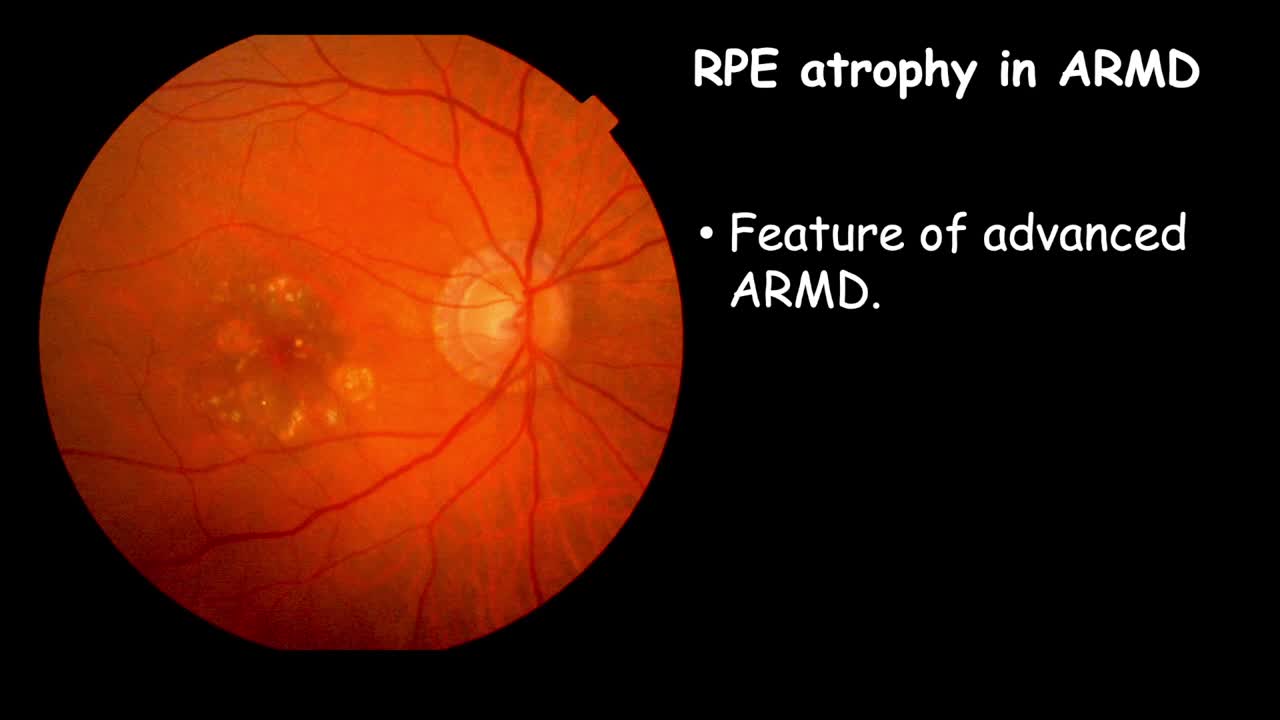
Clinical Changes In Rpe Drusen Eyetube

Find inspiration for Clinical Changes In Rpe Drusen Eyetube with our image finder website, Clinical Changes In Rpe Drusen Eyetube is one of the most popular images and photo galleries in Clinical Changes In Rpe Drusen Eyetube Gallery, Clinical Changes In Rpe Drusen Eyetube Picture are available in collection of high-quality images and discover endless ideas for your living spaces, You will be able to watch high quality photo galleries Clinical Changes In Rpe Drusen Eyetube.
aiartphotoz.com is free images/photos finder and fully automatic search engine, No Images files are hosted on our server, All links and images displayed on our site are automatically indexed by our crawlers, We only help to make it easier for visitors to find a free wallpaper, background Photos, Design Collection, Home Decor and Interior Design photos in some search engines. aiartphotoz.com is not responsible for third party website content. If this picture is your intelectual property (copyright infringement) or child pornography / immature images, please send email to aiophotoz[at]gmail.com for abuse. We will follow up your report/abuse within 24 hours.
Related Images of Clinical Changes In Rpe Drusen Eyetube
Drusen And Degenerative Changes Of The Rpe Cells In Late Onset Macular
Drusen And Degenerative Changes Of The Rpe Cells In Late Onset Macular
520×257
Sd Oct Image Depicting Drusen Types Above The Rpe Sdd A And Below
Sd Oct Image Depicting Drusen Types Above The Rpe Sdd A And Below
724×648
Drusen Rpe Changes And Non Central Ga In A 95 Year Old Patient
Drusen Rpe Changes And Non Central Ga In A 95 Year Old Patient
1280×720
Clinical Changes In Rpe Atrophy Case Report Part Two Eyetube
Clinical Changes In Rpe Atrophy Case Report Part Two Eyetube
678×493
Drusen And Rpe Changes A Colour Fundus Photograph Of The Right Eye
Drusen And Rpe Changes A Colour Fundus Photograph Of The Right Eye
850×1001
Clinical Changes In Rpe Case Report Part Three Youtube
Clinical Changes In Rpe Case Report Part Three Youtube
719×695
An Eye With Neovascularization Associated Rpe Atrophy At The Baseline
An Eye With Neovascularization Associated Rpe Atrophy At The Baseline
611×827
Soft Drusen Underneath A Clearly Defined Rpe Layer Download
Soft Drusen Underneath A Clearly Defined Rpe Layer Download
850×368
Clinical Changes In Rpe Rpe Rip And Atrophy Youtube
Clinical Changes In Rpe Rpe Rip And Atrophy Youtube
600×452
Fundus Image Showing Diffuse Atrophic Retinal Pigment Epithelium Rpe
Fundus Image Showing Diffuse Atrophic Retinal Pigment Epithelium Rpe
640×640
Clinical Retinal Fundus Photograph Of The Right Eye Macula Showing The
Clinical Retinal Fundus Photograph Of The Right Eye Macula Showing The
850×1152
A Representative Image Of Refractile Drusen On Spectral Domain Oct In
A Representative Image Of Refractile Drusen On Spectral Domain Oct In
526×594
Role Of Retinal Pigment Epithelium Encyclopedia Mdpi
Role Of Retinal Pigment Epithelium Encyclopedia Mdpi
1779×1893
Spectrum Of Rpeþbl Disruptions During The Drusenoid Ped Lifecycle
Spectrum Of Rpeþbl Disruptions During The Drusenoid Ped Lifecycle
850×408
Clinical Changes In Rpe Course Ped In Amd Youtube
Clinical Changes In Rpe Course Ped In Amd Youtube
679×247
Refractile Drusen A Near Infrared Reflectance Image Showing
Refractile Drusen A Near Infrared Reflectance Image Showing
1296×728
Drusen In Dense Deposit Disease Not Just Age Related Macular
Drusen In Dense Deposit Disease Not Just Age Related Macular
850×496
Multimodal Imaging Showing Drusen At The Last Clinical Visit All
Multimodal Imaging Showing Drusen At The Last Clinical Visit All
552×144
A Representative Image Of Refractile Drusen On Spectral Domain Oct In
A Representative Image Of Refractile Drusen On Spectral Domain Oct In
850×268
The Clinical Features Of Amd Progression A Cross Section And Fundus
The Clinical Features Of Amd Progression A Cross Section And Fundus
1600×1576
The Left Figure Shows Sd Oct Image Of An Eye With Drusen The Drusen
The Left Figure Shows Sd Oct Image Of An Eye With Drusen The Drusen
638×479
Rpe Demise Linked To The Life Cycle Of Drusenoid Pigment Epithelial
Rpe Demise Linked To The Life Cycle Of Drusenoid Pigment Epithelial
695×276
Clinical Changes In Rpe Atrophy Case Report Part One Eyetube
Clinical Changes In Rpe Atrophy Case Report Part One Eyetube
Optical Coherence Tomography In Age Related Macular Degeneration
Optical Coherence Tomography In Age Related Macular Degeneration
Retinal Pigment Epithelial Rpe Rip The University Of Iowa
Retinal Pigment Epithelial Rpe Rip The University Of Iowa
