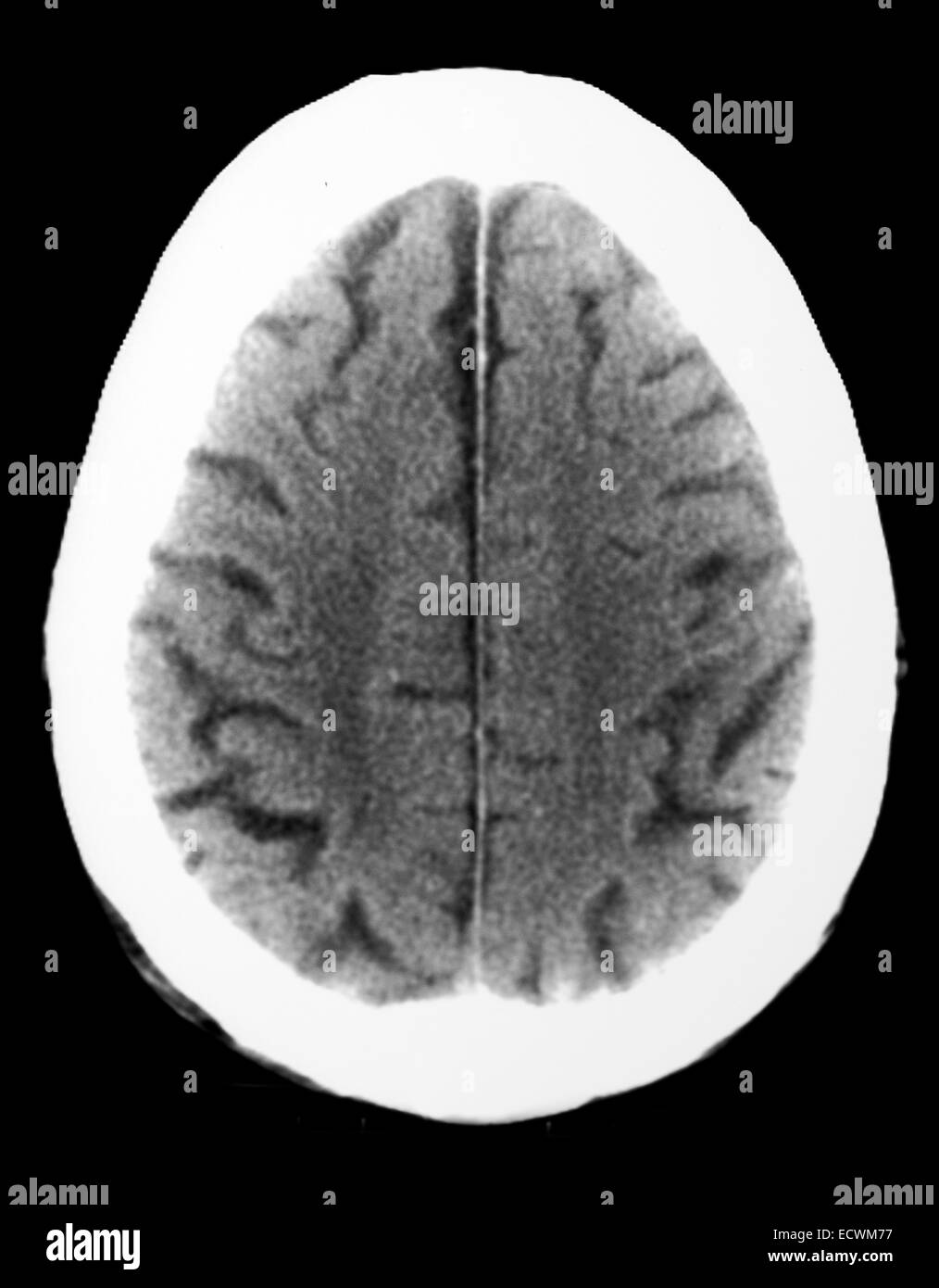
Ct Scan Showing Atrophy Of Both Cerebral Hemispheres Stock Photo Alamy

Find inspiration for Ct Scan Showing Atrophy Of Both Cerebral Hemispheres Stock Photo Alamy with our image finder website, Ct Scan Showing Atrophy Of Both Cerebral Hemispheres Stock Photo Alamy is one of the most popular images and photo galleries in Ct Scan Axial Atrofi Gallery, Ct Scan Showing Atrophy Of Both Cerebral Hemispheres Stock Photo Alamy Picture are available in collection of high-quality images and discover endless ideas for your living spaces, You will be able to watch high quality photo galleries Ct Scan Showing Atrophy Of Both Cerebral Hemispheres Stock Photo Alamy.
aiartphotoz.com is free images/photos finder and fully automatic search engine, No Images files are hosted on our server, All links and images displayed on our site are automatically indexed by our crawlers, We only help to make it easier for visitors to find a free wallpaper, background Photos, Design Collection, Home Decor and Interior Design photos in some search engines. aiartphotoz.com is not responsible for third party website content. If this picture is your intelectual property (copyright infringement) or child pornography / immature images, please send email to aiophotoz[at]gmail.com for abuse. We will follow up your report/abuse within 24 hours.
Related Images of Ct Scan Showing Atrophy Of Both Cerebral Hemispheres Stock Photo Alamy
Ct Scan Showing Atrophy Of Both Cerebral Hemispheres Stock Photo Alamy
Ct Scan Showing Atrophy Of Both Cerebral Hemispheres Stock Photo Alamy
1014×1390
Atrofi Foto Foto Stok Potret And Gambar Bebas Royalti Istock
Atrofi Foto Foto Stok Potret And Gambar Bebas Royalti Istock
612×429
A Muscle Ct Scan Axial Images Of The Thorax Upper Abdomen Lower
A Muscle Ct Scan Axial Images Of The Thorax Upper Abdomen Lower
850×942
Brain Computed Tomography Scan Showing Moderately Severe Atrophy In
Brain Computed Tomography Scan Showing Moderately Severe Atrophy In
850×598
Cureus Role Of Multidetector Computed Tomography In The Evaluation Of
Cureus Role Of Multidetector Computed Tomography In The Evaluation Of
3000×2658
Ct Scan Axial Plane The Defect Width Together With The Lateral Muscles
Ct Scan Axial Plane The Defect Width Together With The Lateral Muscles
640×640
Ct Design And Operation Oncology Medical Physics
Ct Design And Operation Oncology Medical Physics
683×1024
Ketahui Gejala Dan Faktor Penyebab Cerebral Atrophy Terutama Pada Anak
Ketahui Gejala Dan Faktor Penyebab Cerebral Atrophy Terutama Pada Anak
970×544
Cureus Delayed Sub Axial Fracture Dislocation Surgical Management
Cureus Delayed Sub Axial Fracture Dislocation Surgical Management
3000×3550
The Control Axial Ct Scan Through The Right Side Temporal Bone Shows
The Control Axial Ct Scan Through The Right Side Temporal Bone Shows
850×848
Ct Scan Of A Male Patient A Measurement On Axial Ct Scan Demonstrate
Ct Scan Of A Male Patient A Measurement On Axial Ct Scan Demonstrate
850×571
Initial High‐resolution Ct‐scan Axial Lung Window Arrows Indicate
Initial High‐resolution Ct‐scan Axial Lung Window Arrows Indicate
850×1174
Case 1 Neck Contrasted Ct Scan Axial View Showed An Expansive Mass
Case 1 Neck Contrasted Ct Scan Axial View Showed An Expansive Mass
661×1255
Ct Scan Coronal And Axial Axis Showing A Solid Antero Superior
Ct Scan Coronal And Axial Axis Showing A Solid Antero Superior
640×640
Ct Scan Axial Sagittal And Coronal Cuts With 3d Reconstruction Showing
Ct Scan Axial Sagittal And Coronal Cuts With 3d Reconstruction Showing
850×663
Ct Scan Axial Reconstruction Of Right Atrial And Right Ventricular
Ct Scan Axial Reconstruction Of Right Atrial And Right Ventricular
2570×1960
Ct Scan Of Brain Showing Intracerebral Haemorrhage Axial View
Ct Scan Of Brain Showing Intracerebral Haemorrhage Axial View
1596×1172
Ct Scan Step Set Of Upper Body Lung Axial Top View Stock Photo Alamy
Ct Scan Step Set Of Upper Body Lung Axial Top View Stock Photo Alamy
850×1635
Axial Sections Of Abdomen Ct Scan Without Iv Contrast Download
Axial Sections Of Abdomen Ct Scan Without Iv Contrast Download
1247×1390
Axial Scan Chest Hi Res Stock Photography And Images Alamy
Axial Scan Chest Hi Res Stock Photography And Images Alamy
789×643
Ct Scan Shows Bilateral Frontal Brain Atrophy Download Scientific Diagram
Ct Scan Shows Bilateral Frontal Brain Atrophy Download Scientific Diagram
850×231
Ct Scan Axial Coronal And Sagittal Views Download Scientific Diagram
Ct Scan Axial Coronal And Sagittal Views Download Scientific Diagram
850×719
Contrast Enhanced Ct Scan Axial View Shows Enlarged Left Lateral
Contrast Enhanced Ct Scan Axial View Shows Enlarged Left Lateral
850×923
Axial Ct Scan Image Delineation Of The Masticatory Muscles A And
Axial Ct Scan Image Delineation Of The Masticatory Muscles A And
850×675
Axial Sections Of Ct Scan When The Patient First Presented To Our
Axial Sections Of Ct Scan When The Patient First Presented To Our
640×640
Abdominal Ct Scan Axial Image Shows A 10 Cm Dilatation Involving The
Abdominal Ct Scan Axial Image Shows A 10 Cm Dilatation Involving The
615×599
A Computed Tomography Scan Of Brain Showing Cerebral Atrophy And
A Computed Tomography Scan Of Brain Showing Cerebral Atrophy And
804×563
What Are Ct Scans And How Do They Work Live Science
What Are Ct Scans And How Do They Work Live Science
