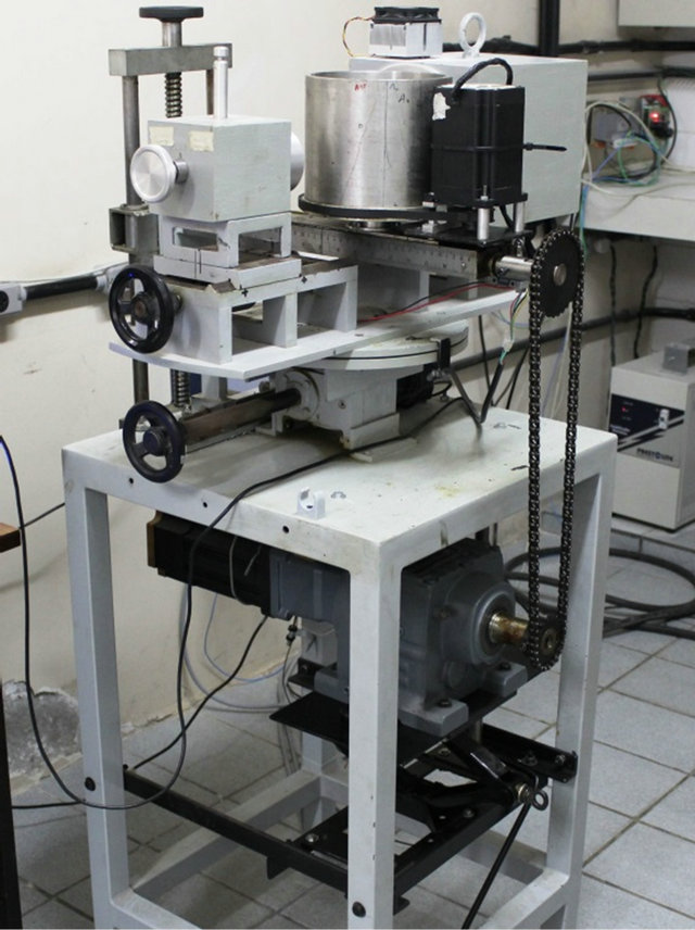
Data Evaluation And Visibility In Single Beam Scanner Of Gamma Ray

Find inspiration for Data Evaluation And Visibility In Single Beam Scanner Of Gamma Ray with our image finder website, Data Evaluation And Visibility In Single Beam Scanner Of Gamma Ray is one of the most popular images and photo galleries in Figure 1 From The Gamma Ray Scanner Of A Chromatographic Plate For Gallery, Data Evaluation And Visibility In Single Beam Scanner Of Gamma Ray Picture are available in collection of high-quality images and discover endless ideas for your living spaces, You will be able to watch high quality photo galleries Data Evaluation And Visibility In Single Beam Scanner Of Gamma Ray.
aiartphotoz.com is free images/photos finder and fully automatic search engine, No Images files are hosted on our server, All links and images displayed on our site are automatically indexed by our crawlers, We only help to make it easier for visitors to find a free wallpaper, background Photos, Design Collection, Home Decor and Interior Design photos in some search engines. aiartphotoz.com is not responsible for third party website content. If this picture is your intelectual property (copyright infringement) or child pornography / immature images, please send email to aiophotoz[at]gmail.com for abuse. We will follow up your report/abuse within 24 hours.
Related Images of Data Evaluation And Visibility In Single Beam Scanner Of Gamma Ray
Figure 1 From The Gamma Ray Scanner Of A Chromatographic Plate For
Figure 1 From The Gamma Ray Scanner Of A Chromatographic Plate For
696×618
Figure 1 From Multiplane Tomographic Gamma Ray Scanner Semantic Scholar
Figure 1 From Multiplane Tomographic Gamma Ray Scanner Semantic Scholar
1654×974
Column Chromatography Made Simple An Easy To Follow Guide
Column Chromatography Made Simple An Easy To Follow Guide
1949×992
Thin Layer Chromatography Tlc Principle And Procedure Owlcation
Thin Layer Chromatography Tlc Principle And Procedure Owlcation
1200×1148
Chromatographic Profile Obtained By Gc Ms Full Scan Of C Nardus Eo
Chromatographic Profile Obtained By Gc Ms Full Scan Of C Nardus Eo
850×417
Schematic Representation For Gamma Ray Attenuation Measurement
Schematic Representation For Gamma Ray Attenuation Measurement
515×515
Data Evaluation And Visibility In Single Beam Scanner Of Gamma Ray
Data Evaluation And Visibility In Single Beam Scanner Of Gamma Ray
640×856
High Performance Liquid Chromatographysize Exclusion Profile Of Gamma
High Performance Liquid Chromatographysize Exclusion Profile Of Gamma
708×680
Photo Of The Dual Source Gamma Ray Computed Tomography Ct Technique
Photo Of The Dual Source Gamma Ray Computed Tomography Ct Technique
850×689
The 32 Channel Gamma Ray Scanner Developed With A Reconstructed
The 32 Channel Gamma Ray Scanner Developed With A Reconstructed
840×240
Figure D1 Gamma Ray Emission Map From Non Thermal Bremsstrahlung
Figure D1 Gamma Ray Emission Map From Non Thermal Bremsstrahlung
800×600
Gamma Ray Spectra Of The Formed Mixture Of Precipitates Packed Into A
Gamma Ray Spectra Of The Formed Mixture Of Precipitates Packed Into A
850×1316
Gamma Ray Spectra Of 15 G 6 Tungstocerateiv Matrix Packed Into A
Gamma Ray Spectra Of 15 G 6 Tungstocerateiv Matrix Packed Into A
850×1158
Schematic Illustration Of The Geometry Of The Mbt Scanner Including
Schematic Illustration Of The Geometry Of The Mbt Scanner Including
850×349
Examples Of The Gamma Ray Spectra Of The Target Material Before Mass
Examples Of The Gamma Ray Spectra Of The Target Material Before Mass
850×327
Upper Panels γ Ray Spectra Coincident With The Mass Peaks Of 89 Rb
Upper Panels γ Ray Spectra Coincident With The Mass Peaks Of 89 Rb
648×893
Gamma Ray Spectra For Blood Samples From The Three Patients Download
Gamma Ray Spectra For Blood Samples From The Three Patients Download
850×1144
High Performance Thin Layer Chromatographic Separation Of α Mangostin
High Performance Thin Layer Chromatographic Separation Of α Mangostin
850×729
Schematic Representation For Gamma Ray Attenuation Measurement
Schematic Representation For Gamma Ray Attenuation Measurement
850×475
Gamma Ray Energy Spectrum Resulting From The Simulation Of Gamma Rays
Gamma Ray Energy Spectrum Resulting From The Simulation Of Gamma Rays
850×618
Figure 1 From Gamma Scanning Data For The Unirradiated Flat Fuel Plates
Figure 1 From Gamma Scanning Data For The Unirradiated Flat Fuel Plates
1088×1608
Gamma Ray And Neutrino Spectra Per Flavor From Agn Coronae With P Inj
Gamma Ray And Neutrino Spectra Per Flavor From Agn Coronae With P Inj
640×640
A Discrete Autocorrelation Function Of The Uppermost 980 M Of Els1
A Discrete Autocorrelation Function Of The Uppermost 980 M Of Els1
850×1818
Gamma Ray Energy Spectrum Resulting From The Simulation Of Gamma Rays
Gamma Ray Energy Spectrum Resulting From The Simulation Of Gamma Rays
640×640
The Gamma Ray By 662 Kev Of Cs Emission Spectra Of Pr Doped Luag
The Gamma Ray By 662 Kev Of Cs Emission Spectra Of Pr Doped Luag
850×675
Gamma Ray Coincidence Spectra For The Three Tsd Bands In 165 Lu The
Gamma Ray Coincidence Spectra For The Three Tsd Bands In 165 Lu The
850×560
Gamma Ray Spectrum Measured With A Naitl Detector At A 60 Cm Position
Gamma Ray Spectrum Measured With A Naitl Detector At A 60 Cm Position
539×382
Gamma Ray Scanner Photos And Premium High Res Pictures Getty Images
Gamma Ray Scanner Photos And Premium High Res Pictures Getty Images
612×435
Gamma Ray Scan Of Healthy Human Lungs Stock Image P5900179 Science
Gamma Ray Scan Of Healthy Human Lungs Stock Image P5900179 Science
800×581
Gamma Ray Spectrum From The Na 22 Source With Solid And Without
Gamma Ray Spectrum From The Na 22 Source With Solid And Without
714×393
Figure 1 From A Decade Of Short Duration Gamma Ray Burst Broad Band
Figure 1 From A Decade Of Short Duration Gamma Ray Burst Broad Band
1418×1152
Secondary Bone Cancer Gamma Scan Stock Image M1340711 Science
Secondary Bone Cancer Gamma Scan Stock Image M1340711 Science
533×800
Male Skeleton Gamma Scan Stock Image P8300046 Science Photo Library
Male Skeleton Gamma Scan Stock Image P8300046 Science Photo Library
800×552
