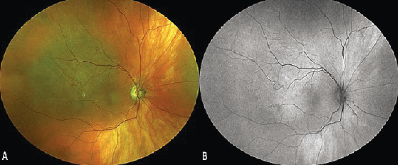
Diagnosing Pigmented Choroidal Lesions

Find inspiration for Diagnosing Pigmented Choroidal Lesions with our image finder website, Diagnosing Pigmented Choroidal Lesions is one of the most popular images and photo galleries in Diagnosing Pigmented Choroidal Lesions Gallery, Diagnosing Pigmented Choroidal Lesions Picture are available in collection of high-quality images and discover endless ideas for your living spaces, You will be able to watch high quality photo galleries Diagnosing Pigmented Choroidal Lesions.
aiartphotoz.com is free images/photos finder and fully automatic search engine, No Images files are hosted on our server, All links and images displayed on our site are automatically indexed by our crawlers, We only help to make it easier for visitors to find a free wallpaper, background Photos, Design Collection, Home Decor and Interior Design photos in some search engines. aiartphotoz.com is not responsible for third party website content. If this picture is your intelectual property (copyright infringement) or child pornography / immature images, please send email to aiophotoz[at]gmail.com for abuse. We will follow up your report/abuse within 24 hours.
Related Images of Diagnosing Pigmented Choroidal Lesions
Figure 1 From Identifying And Treating Pigmented Lesions Of The Choroid
Figure 1 From Identifying And Treating Pigmented Lesions Of The Choroid
1036×752
Figure 1 From Clinical And Spectral Domain Optical Coherence Tomography
Figure 1 From Clinical And Spectral Domain Optical Coherence Tomography
934×1334
Small Pigmented Choroidal Lesion Initially Referred As Nevus A But
Small Pigmented Choroidal Lesion Initially Referred As Nevus A But
662×821
Upper Image Funduscopic Image Of Right Eye With Several Placoid
Upper Image Funduscopic Image Of Right Eye With Several Placoid
600×843
Pigmented Lesions Of The Posterior Eye · • Choroidal Component
Pigmented Lesions Of The Posterior Eye · • Choroidal Component
1200×630
Current Oncology Free Full Text Assessing Choroidal Nevi Melanomas
Current Oncology Free Full Text Assessing Choroidal Nevi Melanomas
2619×1846
Table 1 From Clinical And Spectral Domain Optical Coherence Tomography
Table 1 From Clinical And Spectral Domain Optical Coherence Tomography
934×522
The Ultimate Guide To Diagnosing A Choroidal Nevus
The Ultimate Guide To Diagnosing A Choroidal Nevus
1200×1200
Diagnosing Pigmented Choroidal Review Of Ophthalmology Facebook
Diagnosing Pigmented Choroidal Review Of Ophthalmology Facebook
1080×1080
Mmi Of Choroidal Melanoma Uwf Fundus Image Of A Pigmented Lesion With
Mmi Of Choroidal Melanoma Uwf Fundus Image Of A Pigmented Lesion With
640×640
Changes In Flat Choroidal Lesions Over Time Demonstrated On Enhanced
Changes In Flat Choroidal Lesions Over Time Demonstrated On Enhanced
850×806
Patchy Hypopigmented Subretinal Lesions Involving The Nasal Retinal
Patchy Hypopigmented Subretinal Lesions Involving The Nasal Retinal
850×358
Pigmented Fundus Lesions College Of Optometrists
Pigmented Fundus Lesions College Of Optometrists
445×645
Pigmented Choroidal Lesion A Patient Number 8 In Figure 1 25 Mm
Pigmented Choroidal Lesion A Patient Number 8 In Figure 1 25 Mm
850×333
Pigmented Choroidal Lesions Uhnm Choroidal Naevus Clinic Dr
Pigmented Choroidal Lesions Uhnm Choroidal Naevus Clinic Dr
720×540
A Optomap Composite Image Showing The Typical Appearance Of An
A Optomap Composite Image Showing The Typical Appearance Of An
640×640
