Ear Anatomy Middle Ear
Find inspiration for Ear Anatomy Middle Ear with our image finder website, Ear Anatomy Middle Ear is one of the most popular images and photo galleries in Middle Ear Ct Scan Gallery, Ear Anatomy Middle Ear Picture are available in collection of high-quality images and discover endless ideas for your living spaces, You will be able to watch high quality photo galleries Ear Anatomy Middle Ear.
aiartphotoz.com is free images/photos finder and fully automatic search engine, No Images files are hosted on our server, All links and images displayed on our site are automatically indexed by our crawlers, We only help to make it easier for visitors to find a free wallpaper, background Photos, Design Collection, Home Decor and Interior Design photos in some search engines. aiartphotoz.com is not responsible for third party website content. If this picture is your intelectual property (copyright infringement) or child pornography / immature images, please send email to aiophotoz[at]gmail.com for abuse. We will follow up your report/abuse within 24 hours.
Related Images of Ear Anatomy Middle Ear


Middle Ear Anatomy Ossicles And Tympanic Membrane In Coronal A C
Middle Ear Anatomy Ossicles And Tympanic Membrane In Coronal A C
850×573

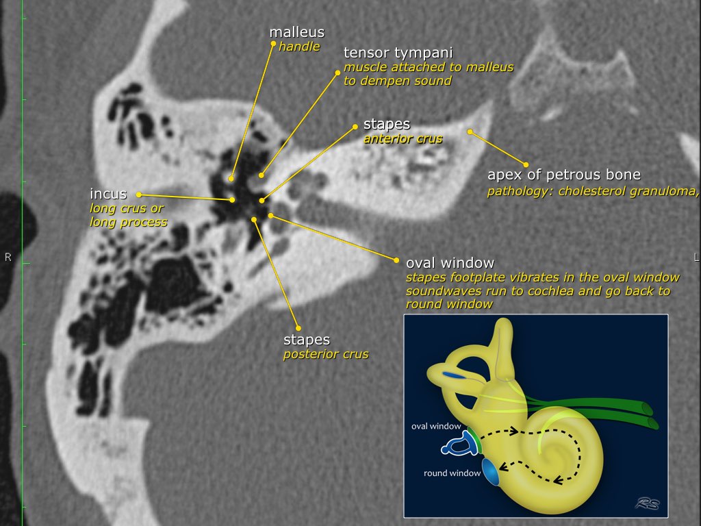
Mastoid Ear Anatomy Anatomy Of The Ear Img Shirely
Mastoid Ear Anatomy Anatomy Of The Ear Img Shirely
1024×1024

Ear Anatomy Ct Scan At Arnulfo Levitt Blog
Ear Anatomy Ct Scan At Arnulfo Levitt Blog
3676×1650

This Illustration Shows The Middle Ear The Tmj And Related
This Illustration Shows The Middle Ear The Tmj And Related
850×632
Pdf Whats Your Diagnosis Symptoms Middle Ear Mass And Unilateral
Pdf Whats Your Diagnosis Symptoms Middle Ear Mass And Unilateral
640×640

Ct Anatomy Of Ear Enteducationswansea
Ct Anatomy Of Ear Enteducationswansea
3677×1648

Ct Scan Of The Middle Ear Anatomy W Radiology
Ct Scan Of The Middle Ear Anatomy W Radiology
400×580

Ct Anatomy Of Ear Enteducationswansea
Ct Anatomy Of Ear Enteducationswansea
3674×1648

Axial Computed Tomography Ct Scans Of The Middle Ear Black Arrows
Axial Computed Tomography Ct Scans Of The Middle Ear Black Arrows
850×753
Ct Scan Of The Middle Ear
Ct Scan Of The Middle Ear
400×580

Middle Ear Anatomy Epitympanum Mesotympanum Hypotympanum Tegmen
Middle Ear Anatomy Epitympanum Mesotympanum Hypotympanum Tegmen
640×640

Axial Horizontal Ct Of The Right Temporal Bone Showing Cholesteatoma
Axial Horizontal Ct Of The Right Temporal Bone Showing Cholesteatoma
850×757

A C Computed Tomography Scan Showing Normal Middle And Inner Ear
A C Computed Tomography Scan Showing Normal Middle And Inner Ear
850×716


Contouring Of The Middle Ear Cavity And The Isthmus Of The Eustachian
Contouring Of The Middle Ear Cavity And The Isthmus Of The Eustachian
717×613

Ear Ct Scan A Axial View The Red Circle Shows The Middle Ear Filled
Ear Ct Scan A Axial View The Red Circle Shows The Middle Ear Filled
640×640

Ct Scan Showing Normal Middle Ear Download Scientific Diagram
Ct Scan Showing Normal Middle Ear Download Scientific Diagram
850×637
Image Of The Patient A And B Pre And Post Operative Ct Scan Images Of
Image Of The Patient A And B Pre And Post Operative Ct Scan Images Of
850×2120
Middle Ear Secretory Otitis A Axial Contrast Enhanced Ct Shows A Mass
Middle Ear Secretory Otitis A Axial Contrast Enhanced Ct Shows A Mass
761×376

Postoperative Imaging Of The Temporal Bone Radiographics
Postoperative Imaging Of The Temporal Bone Radiographics
500×486

Ear Ct Scan A Axial View The Red Circle Shows The Middle Ear Filled
Ear Ct Scan A Axial View The Red Circle Shows The Middle Ear Filled
640×640


Ppt Anatomy And Development Of The Middle Ear Powerpoint Presentation
Ppt Anatomy And Development Of The Middle Ear Powerpoint Presentation
1024×576
A Normal Ear With Middle Ear Volume Of 592 Mm 3 Coronal Ct
A Normal Ear With Middle Ear Volume Of 592 Mm 3 Coronal Ct
850×673

3d Ct Middle And Inner Ear Dr Paulose
3d Ct Middle And Inner Ear Dr Paulose
1024×248

Ct Scan Of The Middle Ear
Ct Scan Of The Middle Ear
400×580
Human Ear 3d Ct Scan Stock Image C0561230 Science Photo Library
Human Ear 3d Ct Scan Stock Image C0561230 Science Photo Library
800×505
