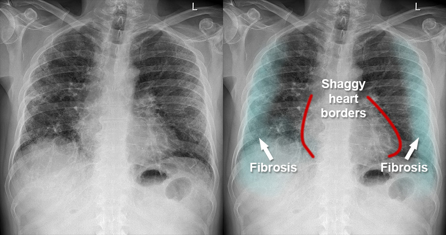
Fibrosis Chest X Ray

Find inspiration for Fibrosis Chest X Ray with our image finder website, Fibrosis Chest X Ray is one of the most popular images and photo galleries in Residual Fibrosis Left Upper Lobe Gallery, Fibrosis Chest X Ray Picture are available in collection of high-quality images and discover endless ideas for your living spaces, You will be able to watch high quality photo galleries Fibrosis Chest X Ray.
aiartphotoz.com is free images/photos finder and fully automatic search engine, No Images files are hosted on our server, All links and images displayed on our site are automatically indexed by our crawlers, We only help to make it easier for visitors to find a free wallpaper, background Photos, Design Collection, Home Decor and Interior Design photos in some search engines. aiartphotoz.com is not responsible for third party website content. If this picture is your intelectual property (copyright infringement) or child pornography / immature images, please send email to aiophotoz[at]gmail.com for abuse. We will follow up your report/abuse within 24 hours.
Related Images of Fibrosis Chest X Ray
All About Pulmonary Fibrosis Residual Fibrosis
All About Pulmonary Fibrosis Residual Fibrosis
703×410
Chest Ct In Case 1 A A Tumor In The Left Upper Lobe Arrow B
Chest Ct In Case 1 A A Tumor In The Left Upper Lobe Arrow B
667×327
Gross Photo Showing Marked Upper Lobe Emphysema With Fibrosis And Lower
Gross Photo Showing Marked Upper Lobe Emphysema With Fibrosis And Lower
429×998
Chest Ct In Case 2 A A Tumor In The Left Upper Lobe Arrow B
Chest Ct In Case 2 A A Tumor In The Left Upper Lobe Arrow B
667×295
Left Upper Lobe Multiple Fibrotic Areas With Cavitation Filled With
Left Upper Lobe Multiple Fibrotic Areas With Cavitation Filled With
850×796
Chest X Ray Showing Multiple Cavitary Lesions Of Left Upper Lobe With
Chest X Ray Showing Multiple Cavitary Lesions Of Left Upper Lobe With
571×749
Case 77 Bilateral Upper Lobe Fibrosis And Pneumothoraces
Case 77 Bilateral Upper Lobe Fibrosis And Pneumothoraces
1687×949
Ct Scan Of The Chest In Lung Window Showing A A Poorly Defined
Ct Scan Of The Chest In Lung Window Showing A A Poorly Defined
850×394
Case 77 Bilateral Upper Lobe Fibrosis And Pneumothoraces
Case 77 Bilateral Upper Lobe Fibrosis And Pneumothoraces
692×389
A Chest X Ray Showed Fibrotic Changes At Both Upper Zones With
A Chest X Ray Showed Fibrotic Changes At Both Upper Zones With
640×640
Cureus Combined Pulmonary Fibrosis And Emphysema And Digital Clubbing
Cureus Combined Pulmonary Fibrosis And Emphysema And Digital Clubbing
3000×1933
And Computed Tomographic Ct Thorax Lung And Mediastinal Sections
And Computed Tomographic Ct Thorax Lung And Mediastinal Sections
800×351
Upper Lobe Sub‐pleural Dense Consolidation And Fibrosis On Computed
Upper Lobe Sub‐pleural Dense Consolidation And Fibrosis On Computed
632×358
Bilateral Upper Lobe Fibrosis With Fissural And Septal Thickening
Bilateral Upper Lobe Fibrosis With Fissural And Septal Thickening
600×492
Coronal Unenhanced Chest Ct Scan Shows Bilateral Fibrotic Upper Lobe
Coronal Unenhanced Chest Ct Scan Shows Bilateral Fibrotic Upper Lobe
850×373
Lung Window Unenhanced Chest Ct Through The Upper A And Mid B Lung
Lung Window Unenhanced Chest Ct Through The Upper A And Mid B Lung
850×690
Chest Ct 1 Year Post Surgery Complete Resolution Re Expansion Of The
Chest Ct 1 Year Post Surgery Complete Resolution Re Expansion Of The
850×461
Fibrotic Pattern Bilateral Sub Pleural Reticulations Arrowheads And
Fibrotic Pattern Bilateral Sub Pleural Reticulations Arrowheads And
736×720
Sarcoidosis With Interstitial Fibrosis And Fungal Ball Left Upper Lobe
Sarcoidosis With Interstitial Fibrosis And Fungal Ball Left Upper Lobe
700×426
Learning Radiology Left Upper Lobe Atelectasis Lul
Learning Radiology Left Upper Lobe Atelectasis Lul
512×610
Chest X Ray Posteroanterior View Right Upper Lobe Pulm Open I
Chest X Ray Posteroanterior View Right Upper Lobe Pulm Open I
729×740
Ct Chest 7272020 Shows A Subpleural Fibrotic Changes With
Ct Chest 7272020 Shows A Subpleural Fibrotic Changes With
768×402
Causes Of Upper Lobe Fibrosis Of The Lung Medical Junction
Causes Of Upper Lobe Fibrosis Of The Lung Medical Junction
1625×1800
Pulmonary Apical Fibrosis In A Patient Treated Earlier For Breast
Pulmonary Apical Fibrosis In A Patient Treated Earlier For Breast
416×577
A 7 Mm Nodule In The Left Upper Lobe Arrow Was Described As A
A 7 Mm Nodule In The Left Upper Lobe Arrow Was Described As A
640×545
