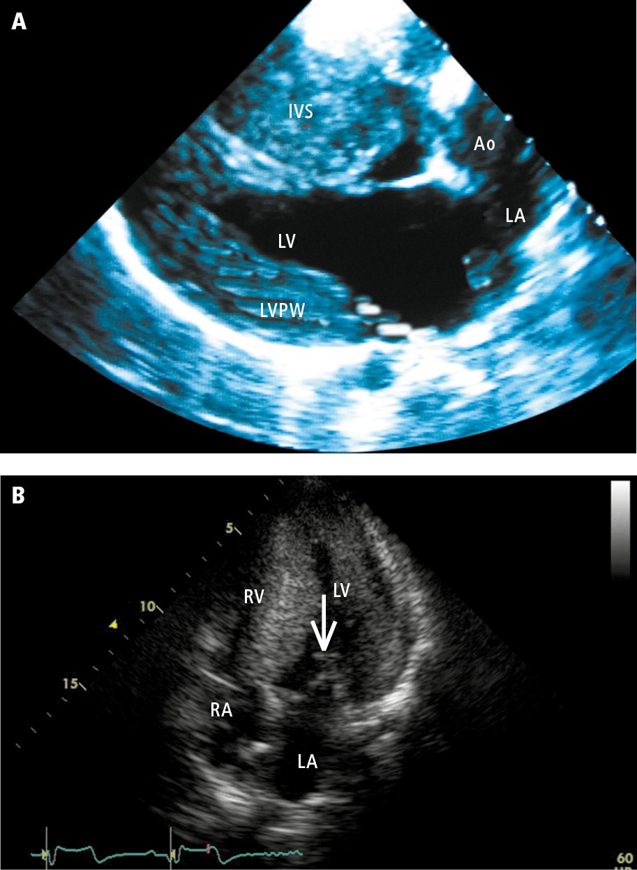
Figure 0310556 Echocardiography Of Patients With Hypertrophic

Find inspiration for Figure 0310556 Echocardiography Of Patients With Hypertrophic with our image finder website, Figure 0310556 Echocardiography Of Patients With Hypertrophic is one of the most popular images and photo galleries in Abnormal Thinning Of The Basal Ventricular Septum A Parasternal Gallery, Figure 0310556 Echocardiography Of Patients With Hypertrophic Picture are available in collection of high-quality images and discover endless ideas for your living spaces, You will be able to watch high quality photo galleries Figure 0310556 Echocardiography Of Patients With Hypertrophic.
aiartphotoz.com is free images/photos finder and fully automatic search engine, No Images files are hosted on our server, All links and images displayed on our site are automatically indexed by our crawlers, We only help to make it easier for visitors to find a free wallpaper, background Photos, Design Collection, Home Decor and Interior Design photos in some search engines. aiartphotoz.com is not responsible for third party website content. If this picture is your intelectual property (copyright infringement) or child pornography / immature images, please send email to aiophotoz[at]gmail.com for abuse. We will follow up your report/abuse within 24 hours.
Related Images of Figure 0310556 Echocardiography Of Patients With Hypertrophic
Abnormal Thinning Of The Basal Ventricular Septum A Parasternal
Abnormal Thinning Of The Basal Ventricular Septum A Parasternal
689×582
Echo Parasternal Long Axis View Revealing Basal Septal Thinning And
Echo Parasternal Long Axis View Revealing Basal Septal Thinning And
703×453
Transthoracic Echocardiography In Parasternal Long Axis View
Transthoracic Echocardiography In Parasternal Long Axis View
850×592
Parasternal Long Axis Of The Left Ventricle Lv Showing The Prominent
Parasternal Long Axis Of The Left Ventricle Lv Showing The Prominent
850×622
Parasternal Long Axis View In A 40 Year Old Woman With Cardiac
Parasternal Long Axis View In A 40 Year Old Woman With Cardiac
704×527
Basal Ventricular Septal Hypertrophy In Systemic Hypertension
Basal Ventricular Septal Hypertrophy In Systemic Hypertension
2500×1371
A Parasternal Long Axis Echocardiographic View At End Diastole
A Parasternal Long Axis Echocardiographic View At End Diastole
850×311
These 3 Cases Demonstrated The Representative Echocardiograms Of
These 3 Cases Demonstrated The Representative Echocardiograms Of
850×288
Basal Septal Bulge In 109 Year Old Man A 2d Parasternal Long Axis View
Basal Septal Bulge In 109 Year Old Man A 2d Parasternal Long Axis View
706×536
Parasternal Long Axis Echocardiogram A Shows The Hypertrophied Basal
Parasternal Long Axis Echocardiogram A Shows The Hypertrophied Basal
850×716
Distinguishing Ventricular Septal Bulge Versus Hypertrophic
Distinguishing Ventricular Septal Bulge Versus Hypertrophic
1315×1800
Figure 0310556 Echocardiography Of Patients With Hypertrophic
Figure 0310556 Echocardiography Of Patients With Hypertrophic
913×1246
Jcm Free Full Text Basal Septal Hypertrophy As The Early Imaging
Jcm Free Full Text Basal Septal Hypertrophy As The Early Imaging
3248×2496
Transthoracic Echocardiography Parasternal Long Axis View With
Transthoracic Echocardiography Parasternal Long Axis View With
635×471
Representative Case Of Evaluation Of Thinning Of The Basal
Representative Case Of Evaluation Of Thinning Of The Basal
640×640
Making Sense Of An Echocardiogram Report For Gps — Cardiology Institute
Making Sense Of An Echocardiogram Report For Gps — Cardiology Institute
991×860
Parasternal Long Axis Echocardiogram Recorded In Diastole A And
Parasternal Long Axis Echocardiogram Recorded In Diastole A And
747×517
Regional Myocardial Contractile Function Wall Motion Abnormalities
Regional Myocardial Contractile Function Wall Motion Abnormalities
1800×1380
The Impact Of Basal Septal Hypertrophy On Outcomes After Transcatheter
The Impact Of Basal Septal Hypertrophy On Outcomes After Transcatheter
1611×1408
Isolated Hypertrophy Of The Basal Ventricular Septum Characteristics
Isolated Hypertrophy Of The Basal Ventricular Septum Characteristics
2370×1138
Transthoracic Echocardiography Parasternal Long Axis View With
Transthoracic Echocardiography Parasternal Long Axis View With
645×472
Imaging Findings At The First Admission A Complete Atrioventricular
Imaging Findings At The First Admission A Complete Atrioventricular
850×801
Parasternal Short Axis View Of The Left Ventricle On Echocardiography
Parasternal Short Axis View Of The Left Ventricle On Echocardiography
459×743
Building Blocks Of The Septal Atrioventricular Junction Region
Building Blocks Of The Septal Atrioventricular Junction Region
834×1116
How To Use Echocardiography To Manage Patients With Shock Medicina
How To Use Echocardiography To Manage Patients With Shock Medicina
3342×2379
Parasternal Short Axis View Papillary Muscle Level Perfusfind
Parasternal Short Axis View Papillary Muscle Level Perfusfind
540×469
Transthoracic Echocardiography Basal Anterior Septum And Posterior
Transthoracic Echocardiography Basal Anterior Septum And Posterior
850×713
Septal Atrioventricular Junction Region Comprehensive Imaging In
Septal Atrioventricular Junction Region Comprehensive Imaging In
1640×1490
Cardiovascular Disease Abnormalities Heart Chambers Risk Factors
Cardiovascular Disease Abnormalities Heart Chambers Risk Factors
1600×1205
Ppt Ventricular Septal Defects Powerpoint Presentation Free Download
Ppt Ventricular Septal Defects Powerpoint Presentation Free Download
1024×768
Apical Muscular Ventricular Septal Defects Between The Left Ventricle
Apical Muscular Ventricular Septal Defects Between The Left Ventricle
1800×1247
