Figure 1 From Hypercementosis And Concrescence Of Maxillary Second
Find inspiration for Figure 1 From Hypercementosis And Concrescence Of Maxillary Second with our image finder website, Figure 1 From Hypercementosis And Concrescence Of Maxillary Second is one of the most popular images and photo galleries in Figure 1 From Hypercementosis And Concrescence Of Maxillary Second Gallery, Figure 1 From Hypercementosis And Concrescence Of Maxillary Second Picture are available in collection of high-quality images and discover endless ideas for your living spaces, You will be able to watch high quality photo galleries Figure 1 From Hypercementosis And Concrescence Of Maxillary Second.
aiartphotoz.com is free images/photos finder and fully automatic search engine, No Images files are hosted on our server, All links and images displayed on our site are automatically indexed by our crawlers, We only help to make it easier for visitors to find a free wallpaper, background Photos, Design Collection, Home Decor and Interior Design photos in some search engines. aiartphotoz.com is not responsible for third party website content. If this picture is your intelectual property (copyright infringement) or child pornography / immature images, please send email to aiophotoz[at]gmail.com for abuse. We will follow up your report/abuse within 24 hours.
Related Images of Figure 1 From Hypercementosis And Concrescence Of Maxillary Second

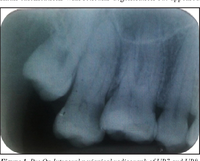
Figure 1 From Hypercementosis And Concrescence Of Maxillary Second
Figure 1 From Hypercementosis And Concrescence Of Maxillary Second
682×550

Figure 1 From Concrescence Of Maxillary Second Molar And Impacted Third
Figure 1 From Concrescence Of Maxillary Second Molar And Impacted Third
756×370

Figure 1 From Hypercementosis And Odontogenic Epithelial Hyperplasia
Figure 1 From Hypercementosis And Odontogenic Epithelial Hyperplasia
1348×550

Figure 1 From A Scanning Electron Microscopic Study Of Hypercementosis
Figure 1 From A Scanning Electron Microscopic Study Of Hypercementosis
646×978
A Panoramic Image Showing Hypercementosis H On The Maxillary Left
A Panoramic Image Showing Hypercementosis H On The Maxillary Left
850×279

Figure 1 From Generalised Hypercementosis A Case Report Semantic Scholar
Figure 1 From Generalised Hypercementosis A Case Report Semantic Scholar
616×400

Figure 1 From Generalised Hypercementosis In A Young Female Seeking
Figure 1 From Generalised Hypercementosis In A Young Female Seeking
692×468

Figure 1 From Hypercementosis A Periscopic Problem Hiding In Plain
Figure 1 From Hypercementosis A Periscopic Problem Hiding In Plain
604×464

Classification Of Roots Type A Simple B Hypercementosis C Thin D
Classification Of Roots Type A Simple B Hypercementosis C Thin D
564×564

Concrescence Of The Right Maxillary Second And Third Molars A Case
Concrescence Of The Right Maxillary Second And Third Molars A Case
2128×1440
Unusual Finding Of Concrescence Bmj Case Reports
Unusual Finding Of Concrescence Bmj Case Reports
1800×1134
Scielo Brasil Concrescence Can The Teeth Involved Be Moved Or
Scielo Brasil Concrescence Can The Teeth Involved Be Moved Or
409×685

Pdf Hypercementosis Diagnostic Imaging By Radiograph Cone Beam
Pdf Hypercementosis Diagnostic Imaging By Radiograph Cone Beam
566×566

Extracted Concrescence Of 17 And 18 A Image Of The Buccal Surface
Extracted Concrescence Of 17 And 18 A Image Of The Buccal Surface
640×640
Figure 1 Presurgical Panoramic Radiograph Showing A Horizontally
Figure 1 Presurgical Panoramic Radiograph Showing A Horizontally
818×392

Figure 2 From Concrescence Of Maxillary Second Molar And Impacted Third
Figure 2 From Concrescence Of Maxillary Second Molar And Impacted Third
996×422

Hypercementosis A Rare Finding In A Patient With Systemic Lupus
Hypercementosis A Rare Finding In A Patient With Systemic Lupus
1800×862

Figure 1 From Endodontic Management Of Three Rooted Maxillary Second
Figure 1 From Endodontic Management Of Three Rooted Maxillary Second
588×1390

Orthopantomograph Showing Abnormal Crown Morphology And Hypercementosis
Orthopantomograph Showing Abnormal Crown Morphology And Hypercementosis
850×455
Figure 3 Specimen Radiograph Showing Concrescent Second And Third
Figure 3 Specimen Radiograph Showing Concrescent Second And Third
460×618

Figure 1 From Fluke Of Fusion And Concrescence In Maxillary Deciduous
Figure 1 From Fluke Of Fusion And Concrescence In Maxillary Deciduous
682×424

Figure 1 From Hypercementosis Associated With Enpp1 Mutations And Gaci
Figure 1 From Hypercementosis Associated With Enpp1 Mutations And Gaci
902×868

Figure 1 From Equine Odontoclastic Tooth Resorption And Hypercementosis
Figure 1 From Equine Odontoclastic Tooth Resorption And Hypercementosis
694×952

Full Article Root Submergence By Decoronation And Root End Resection
Full Article Root Submergence By Decoronation And Root End Resection
500×230

Undiscovered Anatomical Structure In Maxillary Second Molars
Undiscovered Anatomical Structure In Maxillary Second Molars
1772×768


A Intraoral Periapical Radiograph Of The 23242526 Revealing Gross
A Intraoral Periapical Radiograph Of The 23242526 Revealing Gross
716×732

Developmental Alterations Of Tooth Morphology A Root Dilaceration
Developmental Alterations Of Tooth Morphology A Root Dilaceration
566×408

Figure 4 From Endodontic Treatment Of Premolar With Unusual Anatomy And
Figure 4 From Endodontic Treatment Of Premolar With Unusual Anatomy And
532×1502

Concrescence Of Impacted Mandibular Third Molar With A Fourth Molar
Concrescence Of Impacted Mandibular Third Molar With A Fourth Molar
660×298


Diagnosing Oral And Maxillofacial Pathologies In My Residency
Diagnosing Oral And Maxillofacial Pathologies In My Residency
1600×1033


Figure 1 From Hypercementosis Associated With Enpp1 Mutations And Gaci
Figure 1 From Hypercementosis Associated With Enpp1 Mutations And Gaci
886×784

Opg Showing Hypercementosis In 14 And 15 Regions Download Scientific
Opg Showing Hypercementosis In 14 And 15 Regions Download Scientific
668×330
