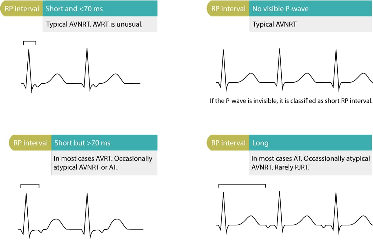
Figure X Differential Diagnoses Based On Rp Interval Ecg Learning

Find inspiration for Figure X Differential Diagnoses Based On Rp Interval Ecg Learning with our image finder website, Figure X Differential Diagnoses Based On Rp Interval Ecg Learning is one of the most popular images and photo galleries in Figure X Differential Diagnoses Based On Rp Interval Ecg Learning Gallery, Figure X Differential Diagnoses Based On Rp Interval Ecg Learning Picture are available in collection of high-quality images and discover endless ideas for your living spaces, You will be able to watch high quality photo galleries Figure X Differential Diagnoses Based On Rp Interval Ecg Learning.
aiartphotoz.com is free images/photos finder and fully automatic search engine, No Images files are hosted on our server, All links and images displayed on our site are automatically indexed by our crawlers, We only help to make it easier for visitors to find a free wallpaper, background Photos, Design Collection, Home Decor and Interior Design photos in some search engines. aiartphotoz.com is not responsible for third party website content. If this picture is your intelectual property (copyright infringement) or child pornography / immature images, please send email to aiophotoz[at]gmail.com for abuse. We will follow up your report/abuse within 24 hours.
Related Images of Figure X Differential Diagnoses Based On Rp Interval Ecg Learning
Figure X Differential Diagnoses Based On Rp Interval Ecg Learning
Figure X Differential Diagnoses Based On Rp Interval Ecg Learning
1200×774
Schematic Illustration Of Rp Interval Measurement And Classification
Schematic Illustration Of Rp Interval Measurement And Classification
736×831
Ecg Rhythms Differential Diagnosis Of Long Rp Tachycardia With
Ecg Rhythms Differential Diagnosis Of Long Rp Tachycardia With
1600×873
Ppt Tachyarrhythmia Pearls For Ecg Diagnosis Powerpoint Presentation
Ppt Tachyarrhythmia Pearls For Ecg Diagnosis Powerpoint Presentation
1024×768
Supraventricular Tachycardia Electrocardiogram Diagnosis And Clinical
Supraventricular Tachycardia Electrocardiogram Diagnosis And Clinical
482×621
Schematic Of Ecg Indicating The Rr Interval Variation Download
Schematic Of Ecg Indicating The Rr Interval Variation Download
850×237
A Long Rp Tachycardia Where Is The Culprit Heart Rhythm
A Long Rp Tachycardia Where Is The Culprit Heart Rhythm
3155×1780
St Segment Elevation In Acute Myocardial Ischemia And Differential
St Segment Elevation In Acute Myocardial Ischemia And Differential
1300×957
Long Rp Supraventricular Tachycardia What Is The Mechanism Heart Rhythm
Long Rp Supraventricular Tachycardia What Is The Mechanism Heart Rhythm
1642×991
Pacemaker Refractory Periods Rp This Ecg Demonstrates The Timings Of
Pacemaker Refractory Periods Rp This Ecg Demonstrates The Timings Of
734×260
A Long Rp Supraventricular Tachycardia With The Earliest Atrial
A Long Rp Supraventricular Tachycardia With The Earliest Atrial
581×429
R Wave Qrs Complex And Rr Interval In Standard Ecg Signals Download
R Wave Qrs Complex And Rr Interval In Standard Ecg Signals Download
663×374
Differential Diagnoses T Wave Inversions And Prolonged Qtc Interval
Differential Diagnoses T Wave Inversions And Prolonged Qtc Interval
1200×838
A Patients Presenting Rhythm A Narrow Complex Short Rp Tachycardia
A Patients Presenting Rhythm A Narrow Complex Short Rp Tachycardia
850×528
Diagnosis And Management Of Narrow And Wide Complex Tachycardia
Diagnosis And Management Of Narrow And Wide Complex Tachycardia
768×810
Figure 3 Differential Diagnoses In St Segment Depressions Ecg Learning
Figure 3 Differential Diagnoses In St Segment Depressions Ecg Learning
1080×1266
Supraventricular Arrhythmias Clinical Framework And Common Scenarios
Supraventricular Arrhythmias Clinical Framework And Common Scenarios
1761×1462
Long Rp Supraventricular Tachycardia What Is The Mechanism Heart Rhythm
Long Rp Supraventricular Tachycardia What Is The Mechanism Heart Rhythm
1642×867
St Segment Elevation In Acute Myocardial Ischemia And Differential
St Segment Elevation In Acute Myocardial Ischemia And Differential
1000×693
An Approach To Diagnosing Supraventricular Tachycardias On The 12 Lead
An Approach To Diagnosing Supraventricular Tachycardias On The 12 Lead
679×446
Atrioventricular Nodal Reentry Tachycardia Avnrt Ecg Features
Atrioventricular Nodal Reentry Tachycardia Avnrt Ecg Features
1200×1541
Figure 1 The Qt Interval On The Ecg Ecg Learning
Figure 1 The Qt Interval On The Ecg Ecg Learning
700×560
J Point Ecg Interval • Litfl Medical Blog • Ecg Library Basics
J Point Ecg Interval • Litfl Medical Blog • Ecg Library Basics
1000×601
Ecg In Left Ventricular Hypertrophy Lvh Criteria And Implications
Ecg In Left Ventricular Hypertrophy Lvh Criteria And Implications
1200×1413
Approach To Patients With Chest Pain Differential Diagnoses
Approach To Patients With Chest Pain Differential Diagnoses
1500×2003
How To Interpret The Ecg Ekg A Systematic Approach Ecg Learning
How To Interpret The Ecg Ekg A Systematic Approach Ecg Learning
1400×904
