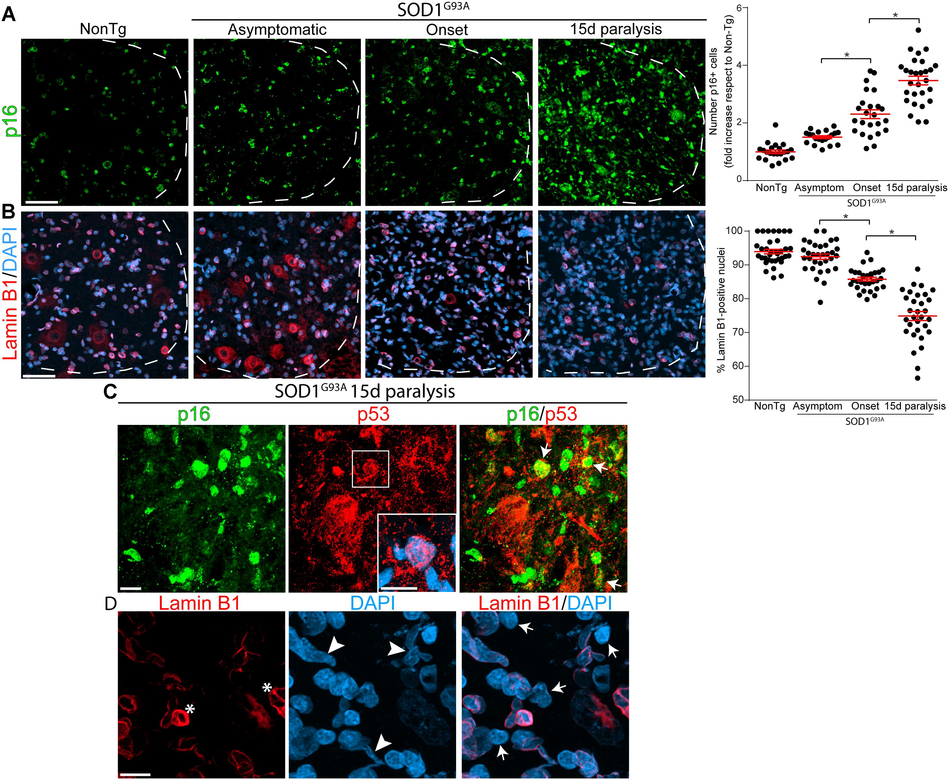
Frontiers Emergence Of Microglia Bearing Senescence Markers During

Find inspiration for Frontiers Emergence Of Microglia Bearing Senescence Markers During with our image finder website, Frontiers Emergence Of Microglia Bearing Senescence Markers During is one of the most popular images and photo galleries in Figure 1 From Dynamic Responses Of Microglia In Animal Models Of Gallery, Frontiers Emergence Of Microglia Bearing Senescence Markers During Picture are available in collection of high-quality images and discover endless ideas for your living spaces, You will be able to watch high quality photo galleries Frontiers Emergence Of Microglia Bearing Senescence Markers During.
aiartphotoz.com is free images/photos finder and fully automatic search engine, No Images files are hosted on our server, All links and images displayed on our site are automatically indexed by our crawlers, We only help to make it easier for visitors to find a free wallpaper, background Photos, Design Collection, Home Decor and Interior Design photos in some search engines. aiartphotoz.com is not responsible for third party website content. If this picture is your intelectual property (copyright infringement) or child pornography / immature images, please send email to aiophotoz[at]gmail.com for abuse. We will follow up your report/abuse within 24 hours.
Related Images of Frontiers Emergence Of Microglia Bearing Senescence Markers During
Figure 1 From Dynamic Responses Of Microglia In Animal Models Of
Figure 1 From Dynamic Responses Of Microglia In Animal Models Of
1092×628
Dynamic Responses Of Microglia In Animal Models Of Multiple Sclerosis
Dynamic Responses Of Microglia In Animal Models Of Multiple Sclerosis
797×559
Figure 1 From Microglia As Dynamic Cellular Mediators Of Brain Function
Figure 1 From Microglia As Dynamic Cellular Mediators Of Brain Function
1002×886
Figure 1 From The Pathophysiological Role Of Microglia In Dynamic
Figure 1 From The Pathophysiological Role Of Microglia In Dynamic
1168×724
Figure 1 From Dynamic Responses Of Microglia In Animal Models Of
Figure 1 From Dynamic Responses Of Microglia In Animal Models Of
1166×664
Schematic Of The Potential Mechanisms Of Glp 1r In The Tnc In The Cm
Schematic Of The Potential Mechanisms Of Glp 1r In The Tnc In The Cm
850×682
Frontiers Microglia Immune Regulators Of Neurodevelopment
Frontiers Microglia Immune Regulators Of Neurodevelopment
510×733
Pdf Dynamic Responses Of Microglia In Animal Models Of Multiple Sclerosis
Pdf Dynamic Responses Of Microglia In Animal Models Of Multiple Sclerosis
850×1113
Figure 1 From Explorer A Zebrafish Live Imaging Model Reveals
Figure 1 From Explorer A Zebrafish Live Imaging Model Reveals
1004×588
Schematic Of Microglia Activation And Responses In The Cuprizone
Schematic Of Microglia Activation And Responses In The Cuprizone
640×640
Figure 1 From Microglia And The Control Of Autoreactive T Cell
Figure 1 From Microglia And The Control Of Autoreactive T Cell
802×736
Figure 1 From The Dynamic Role Of Microglia And The Endocannabinoid
Figure 1 From The Dynamic Role Of Microglia And The Endocannabinoid
934×756
Microglia Are Phenotypically Dynamic A The Functional Roles Of
Microglia Are Phenotypically Dynamic A The Functional Roles Of
640×640
Physiological Function Of Microglia A Microglia Constantly Monitor
Physiological Function Of Microglia A Microglia Constantly Monitor
850×822
Structuraldynamic Interactions Between Microglia And Dendritic Spines
Structuraldynamic Interactions Between Microglia And Dendritic Spines
850×820
Frontiers A Comparative Biology Of Microglia Across Species
Frontiers A Comparative Biology Of Microglia Across Species
2085×1026
Figure 1 From Exosomes From Microglia Attenuate Photoreceptor Injury
Figure 1 From Exosomes From Microglia Attenuate Photoreceptor Injury
662×1034
Frontiers Aged Microglia In Neurodegenerative Diseases Microglia
Frontiers Aged Microglia In Neurodegenerative Diseases Microglia
4550×3206
Frontiers Microglial Phenotypic Transition Signaling Pathways And
Frontiers Microglial Phenotypic Transition Signaling Pathways And
3850×2254
Fifty Shades Of Microglia Trends In Neurosciences
Fifty Shades Of Microglia Trends In Neurosciences
2590×3354
Frontiers Microglia Dynamic Response And Phenotype Heterogeneity In
Frontiers Microglia Dynamic Response And Phenotype Heterogeneity In
1784×2363
Schematic Of The Long Lasting Activation And Of The Dynamic Changes Of
Schematic Of The Long Lasting Activation And Of The Dynamic Changes Of
656×903
Human Microglia States Are Conserved Across Experimental Models And
Human Microglia States Are Conserved Across Experimental Models And
2906×2681
Frontiers Microglia The Hub Of Intercellular Communication In
Frontiers Microglia The Hub Of Intercellular Communication In
1300×733
Frontiers The Role Of Microglia In Bacterial Meningitis Inflammatory
Frontiers The Role Of Microglia In Bacterial Meningitis Inflammatory
6017×4457
Human Microglia States Are Conserved Across Experimental Models And
Human Microglia States Are Conserved Across Experimental Models And
3321×2861
Sexual Dimorphism Of Microglia And Synapses During Mouse Postnatal
Sexual Dimorphism Of Microglia And Synapses During Mouse Postnatal
1129×852
Frontiers Overview Of General And Discriminating Markers Of
Frontiers Overview Of General And Discriminating Markers Of
1300×946
Prenatal Inflammation Shapes Microglial Immune Response Into Adulthood
Prenatal Inflammation Shapes Microglial Immune Response Into Adulthood
3389×1882
Frontiers Role Of Microglia In Brain Development After Viral Infection
Frontiers Role Of Microglia In Brain Development After Viral Infection
1065×654
Frontiers Emergence Of Microglia Bearing Senescence Markers During
Frontiers Emergence Of Microglia Bearing Senescence Markers During
3015×2480
Table 1 From Animal Models Of Ms Reveal Multiple Roles Of Microglia In
Table 1 From Animal Models Of Ms Reveal Multiple Roles Of Microglia In
1400×1064
Figures And Data In The Role Of Microglia And Their Cx3cr1 Signaling In
Figures And Data In The Role Of Microglia And Their Cx3cr1 Signaling In
617×546
Model Of How Microglia Activation Can Be Specifically Controlled By
Model Of How Microglia Activation Can Be Specifically Controlled By
850×432
Three Step Model Of Microglial Phagocytosis In Physiological
Three Step Model Of Microglial Phagocytosis In Physiological
843×254
