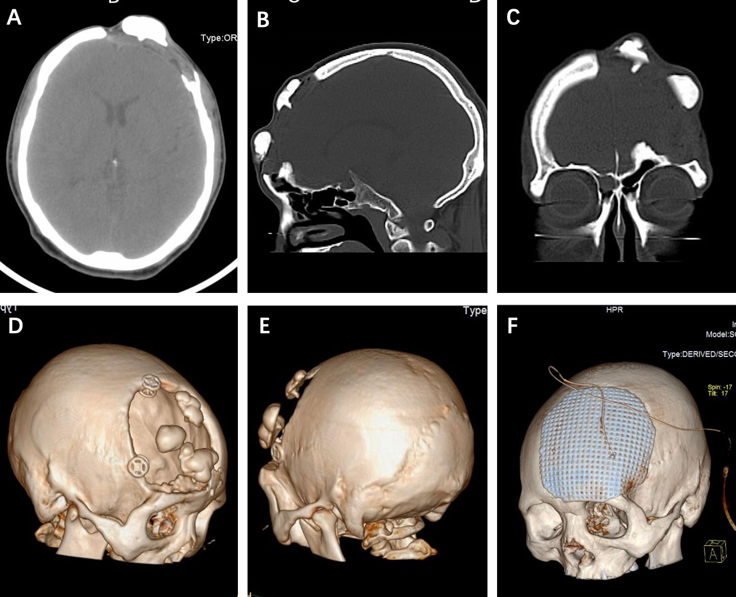
Frontiers Relapse Of Skull Osteoma After Hydroxyapatite Cement

Find inspiration for Frontiers Relapse Of Skull Osteoma After Hydroxyapatite Cement with our image finder website, Frontiers Relapse Of Skull Osteoma After Hydroxyapatite Cement is one of the most popular images and photo galleries in Clinical Image Showing Abnormal Skull Shape With Steeply Rising Gallery, Frontiers Relapse Of Skull Osteoma After Hydroxyapatite Cement Picture are available in collection of high-quality images and discover endless ideas for your living spaces, You will be able to watch high quality photo galleries Frontiers Relapse Of Skull Osteoma After Hydroxyapatite Cement.
aiartphotoz.com is free images/photos finder and fully automatic search engine, No Images files are hosted on our server, All links and images displayed on our site are automatically indexed by our crawlers, We only help to make it easier for visitors to find a free wallpaper, background Photos, Design Collection, Home Decor and Interior Design photos in some search engines. aiartphotoz.com is not responsible for third party website content. If this picture is your intelectual property (copyright infringement) or child pornography / immature images, please send email to aiophotoz[at]gmail.com for abuse. We will follow up your report/abuse within 24 hours.
Related Images of Frontiers Relapse Of Skull Osteoma After Hydroxyapatite Cement
Clinical Image Showing Abnormal Skull Shape With Steeply Rising
Clinical Image Showing Abnormal Skull Shape With Steeply Rising
850×394
Craniosynostosis Selected Craniofacial Syndromes And Other
Craniosynostosis Selected Craniofacial Syndromes And Other
650×716
Craniofacial Anomalies Facial Plastic Surgery Clinics
Craniofacial Anomalies Facial Plastic Surgery Clinics
2856×4143
Congenital Anomalies Of The Skull Clinical Gate
Congenital Anomalies Of The Skull Clinical Gate
650×313
Frontiers Relapse Of Skull Osteoma After Hydroxyapatite Cement
Frontiers Relapse Of Skull Osteoma After Hydroxyapatite Cement
1454×1180
Racgp Paediatric Head Shape And Craniosynostosis
Racgp Paediatric Head Shape And Craniosynostosis
760×662
3 D Ct Scans Of Infant With Deformed Skull Stock Image M3500091
3 D Ct Scans Of Infant With Deformed Skull Stock Image M3500091
771×800
Case 3 Three Dimensional Reformatted Ct Scan Imaging Demonstrates Head
Case 3 Three Dimensional Reformatted Ct Scan Imaging Demonstrates Head
850×906
A Shows Normal B Abnormal Skull With Ccd Download Scientific Diagram
A Shows Normal B Abnormal Skull With Ccd Download Scientific Diagram
767×425
Abnormal Skull Shape Craniofacial Surgery Cranial Vault Surgery
Abnormal Skull Shape Craniofacial Surgery Cranial Vault Surgery
578×692
Characteristics Of Normal And Abnormal Postnatal Craniofacial Growth
Characteristics Of Normal And Abnormal Postnatal Craniofacial Growth
680×432
Pediatric Craniosynostosis Uf Pediatric Neurosurgery Pediatric
Pediatric Craniosynostosis Uf Pediatric Neurosurgery Pediatric
727×735
Lateral Skull Radiograph Of First Patient Shows Asymmetrical Calvarial
Lateral Skull Radiograph Of First Patient Shows Asymmetrical Calvarial
720×482
Understanding Craniosynostosis Causes Symptoms Complications And
Understanding Craniosynostosis Causes Symptoms Complications And
675×328
The Skull X Ray Findings The Abnormal Calcifications Arrows Were
The Skull X Ray Findings The Abnormal Calcifications Arrows Were
600×466
X Ray Of Skull Fracture X Ray Of Skull Fractureabnormal Skull Xray
X Ray Of Skull Fracture X Ray Of Skull Fractureabnormal Skull Xray
609×517
A 59 Year Old Female With Back Pain And An Abnormal Bone Scan That
A 59 Year Old Female With Back Pain And An Abnormal Bone Scan That
650×662
Skull 3 Upper Row Gross Morphology In Left Lateral Views Showing
Skull 3 Upper Row Gross Morphology In Left Lateral Views Showing
418×500
Frontiers A Large Skull Defect Due To Gorham Stout Disease Case
Frontiers A Large Skull Defect Due To Gorham Stout Disease Case
726×744
Clinical Photograph Of First Patient Showing Abnormal Shape Of Head
Clinical Photograph Of First Patient Showing Abnormal Shape Of Head
640×640
Skull Radiography Lateral View Showing Copper Beaten Appearance With
Skull Radiography Lateral View Showing Copper Beaten Appearance With
689×947
A Clinical Picture Of The Proband Showing Features Of Microcephaly
A Clinical Picture Of The Proband Showing Features Of Microcephaly
640×640
Clinical Case 1 Postmortem X Ray Of The Head Normal Shape And Size Of
Clinical Case 1 Postmortem X Ray Of The Head Normal Shape And Size Of
850×804
Clinical Image Of The Patient Showing Acrogigantism Height 192 Cm A
Clinical Image Of The Patient Showing Acrogigantism Height 192 Cm A
1802×1374
Imaging Spectrum Of Calvarial Abnormalities Radiographics
Imaging Spectrum Of Calvarial Abnormalities Radiographics
598×425
Reconstruction Image Of Computer Tomography Of The Abnormal Skull
Reconstruction Image Of Computer Tomography Of The Abnormal Skull
3600×2320
Craniosynostosis Understanding The Misshaped Head Radiographics
Craniosynostosis Understanding The Misshaped Head Radiographics
600×459
Abnormal Skull Shape Observed By Conventional Sonographic Scan
Abnormal Skull Shape Observed By Conventional Sonographic Scan
576×735
