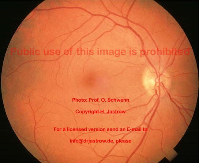
Human Retina Overview Clinical Anatomy

Find inspiration for Human Retina Overview Clinical Anatomy with our image finder website, Human Retina Overview Clinical Anatomy is one of the most popular images and photo galleries in Retina Excavation Of The Papilla Right Eye A Photo On Flickriver Gallery, Human Retina Overview Clinical Anatomy Picture are available in collection of high-quality images and discover endless ideas for your living spaces, You will be able to watch high quality photo galleries Human Retina Overview Clinical Anatomy.
aiartphotoz.com is free images/photos finder and fully automatic search engine, No Images files are hosted on our server, All links and images displayed on our site are automatically indexed by our crawlers, We only help to make it easier for visitors to find a free wallpaper, background Photos, Design Collection, Home Decor and Interior Design photos in some search engines. aiartphotoz.com is not responsible for third party website content. If this picture is your intelectual property (copyright infringement) or child pornography / immature images, please send email to aiophotoz[at]gmail.com for abuse. We will follow up your report/abuse within 24 hours.
Related Images of Human Retina Overview Clinical Anatomy
Retina Excavation Of The Papilla Right Eye A Photo On Flickriver
Retina Excavation Of The Papilla Right Eye A Photo On Flickriver
1024×819
¿qué Es La Excavación Papilar Y Por Qué Ocurre Área Oftalmológica
¿qué Es La Excavación Papilar Y Por Qué Ocurre Área Oftalmológica
1200×900
Retinography Showing A White Atrophic Papilla With Calcified Deposits
Retinography Showing A White Atrophic Papilla With Calcified Deposits
850×848
Excavation With Retinal Pigment Epithelium Rpe Changes These Images
Excavation With Retinal Pigment Epithelium Rpe Changes These Images
600×600
Subfoveal Focal Choroidal Excavation With Macular Vortex Vein
Subfoveal Focal Choroidal Excavation With Macular Vortex Vein
2173×2174
Color Fundus A Of The Right Eye Shows A White Area Of Excavation In
Color Fundus A Of The Right Eye Shows A White Area Of Excavation In
850×375
Fundus Examination Of Both Eyes And Sd Oct Scan A Right Eye Re
Fundus Examination Of Both Eyes And Sd Oct Scan A Right Eye Re
850×844
Ophthalmoscopy Papilla Discretely Tilted Medial Pale Pink And Well
Ophthalmoscopy Papilla Discretely Tilted Medial Pale Pink And Well
850×435
Case 2 A Fundus Image Of The Right Eye Shows A Deep Excavation Of
Case 2 A Fundus Image Of The Right Eye Shows A Deep Excavation Of
558×637
Large Anomalous Optic Disc With Conical Excavation Significant
Large Anomalous Optic Disc With Conical Excavation Significant
660×551
A Re Well Defined And Not Over Elevated Papilla Inferior And
A Re Well Defined And Not Over Elevated Papilla Inferior And
640×640
Figure1funduscopic Examination Papilledema Retinal Hemorrhage Soft
Figure1funduscopic Examination Papilledema Retinal Hemorrhage Soft
827×663
Correlation Between Focal Choroidal Excavation And Underlyin Retina
Correlation Between Focal Choroidal Excavation And Underlyin Retina
1200×1339
Findings In A 58 Year Old Man With Bilateral Focal Choroidal
Findings In A 58 Year Old Man With Bilateral Focal Choroidal
787×714
A Fundoscopy Right Eye Ill Defined Greyish Retinal Mass On The
A Fundoscopy Right Eye Ill Defined Greyish Retinal Mass On The
850×375
Valuing The Eyes Nikon Corporation Healthcare Business Unit
Valuing The Eyes Nikon Corporation Healthcare Business Unit
936×586
Focal Choroidal Excavation December 2018 Illinois Retina Associates
Focal Choroidal Excavation December 2018 Illinois Retina Associates
1397×1084
Peripapillary Focal Choroidal Excavation In Association With Retina
Peripapillary Focal Choroidal Excavation In Association With Retina
1200×423
Peripapillary Staphyloma Cataract And Other Lens Disorders Jama
Peripapillary Staphyloma Cataract And Other Lens Disorders Jama
520×430
Decompression Retinopathy Retinal Hemorrhages Papilla And Macular
Decompression Retinopathy Retinal Hemorrhages Papilla And Macular
680×682
Atlas Entry Peripapillary Combined Hamartoma Of The Retina And
Atlas Entry Peripapillary Combined Hamartoma Of The Retina And
686×720
Focal Choroidal Excavation Review Of Literature British Journal Of
Focal Choroidal Excavation Review Of Literature British Journal Of
1280×828
The Expanded Spectrum Of Focal Choroidal Excavation Ophthalmic
The Expanded Spectrum Of Focal Choroidal Excavation Ophthalmic
520×316
Le Fond Doeil éclairé à Lophtalmoscope Ophtalmologie
Le Fond Doeil éclairé à Lophtalmoscope Ophtalmologie
500×322
Observation N° 2 Fond Doeil Excavation Papillaire Bilatérale Plus
Observation N° 2 Fond Doeil Excavation Papillaire Bilatérale Plus
689×289
Excavación Papilar ¿qué Es Y Cuáles Son Sus Causas Blog De Clínica
Excavación Papilar ¿qué Es Y Cuáles Son Sus Causas Blog De Clínica
1037×1011
