Hypoechoic Liver Lobe Case Studies
Find inspiration for Hypoechoic Liver Lobe Case Studies with our image finder website, Hypoechoic Liver Lobe Case Studies is one of the most popular images and photo galleries in Hypoechoic Liver Lobe Case Studies Gallery, Hypoechoic Liver Lobe Case Studies Picture are available in collection of high-quality images and discover endless ideas for your living spaces, You will be able to watch high quality photo galleries Hypoechoic Liver Lobe Case Studies.
aiartphotoz.com is free images/photos finder and fully automatic search engine, No Images files are hosted on our server, All links and images displayed on our site are automatically indexed by our crawlers, We only help to make it easier for visitors to find a free wallpaper, background Photos, Design Collection, Home Decor and Interior Design photos in some search engines. aiartphotoz.com is not responsible for third party website content. If this picture is your intelectual property (copyright infringement) or child pornography / immature images, please send email to aiophotoz[at]gmail.com for abuse. We will follow up your report/abuse within 24 hours.
Related Images of Hypoechoic Liver Lobe Case Studies


Ct Scan Revealed Hypoechoic Lesion In The Right Lobe Of The Liver
Ct Scan Revealed Hypoechoic Lesion In The Right Lobe Of The Liver
850×475
A Slightly Hypoechoic Fll In The Right Liver Lobe B The Fll Shows
A Slightly Hypoechoic Fll In The Right Liver Lobe B The Fll Shows
850×443

Liver Ultrasound Shows Two Heterogeneous Hypoechoic Lesions With
Liver Ultrasound Shows Two Heterogeneous Hypoechoic Lesions With
720×387
Imaging Studies A Abdominal Ultrasonography Revealed Irregular
Imaging Studies A Abdominal Ultrasonography Revealed Irregular
850×633

Ultrasound Showed Multiple Hypoechoic Nodules In The Liver And Spleen
Ultrasound Showed Multiple Hypoechoic Nodules In The Liver And Spleen
600×299

Large Hypoechoic Nodular Lesions In Liver Right Lobe Detected By
Large Hypoechoic Nodular Lesions In Liver Right Lobe Detected By
850×990
A Hypoechoic Nodule In Segment Vii Of A Non Cirrhotic Liver Normal
A Hypoechoic Nodule In Segment Vii Of A Non Cirrhotic Liver Normal
645×1193

Usg Abdomen Showing Hypoechoic Lesions In Right Lobe Of Liver Largest
Usg Abdomen Showing Hypoechoic Lesions In Right Lobe Of Liver Largest
758×567

Usceus Examination A Hypoechoic Mass Of Right Lobe Of Liver B The
Usceus Examination A Hypoechoic Mass Of Right Lobe Of Liver B The
850×253
A Slightly Hypoechoic Fll In The Right Liver Lobe B The Fll Shows
A Slightly Hypoechoic Fll In The Right Liver Lobe B The Fll Shows
640×640
A B Mode Ultrasound With Hypoechoic Liver Lesions In The Right Liver
A B Mode Ultrasound With Hypoechoic Liver Lesions In The Right Liver
640×640

Homogonous Hypoechoic Lesion Anterior To The Left Hepatic Lobe Color
Homogonous Hypoechoic Lesion Anterior To The Left Hepatic Lobe Color
640×640

Two Hypoechoic Round Lesions In The Left Lobe Of The Liver Download
Two Hypoechoic Round Lesions In The Left Lobe Of The Liver Download
660×475

A Sonography Of The Liver Shows Hypoechoic Appearance Over Caudate
A Sonography Of The Liver Shows Hypoechoic Appearance Over Caudate
850×619

Well Defined Hypoechoic Lesion In Liver Segment Viii B First
Well Defined Hypoechoic Lesion In Liver Segment Viii B First
850×340

Large Hypoechoic Non Homogeneous Mass In The Right Lobe Of The Liver
Large Hypoechoic Non Homogeneous Mass In The Right Lobe Of The Liver
850×389

Ultrasound Shows Hypoechoic Nodule In The Hepatic Lobe And Cdfi Shows
Ultrasound Shows Hypoechoic Nodule In The Hepatic Lobe And Cdfi Shows
600×299

Usceus Examination A Hypoechoic Mass Of Right Lobe Of Liver B The
Usceus Examination A Hypoechoic Mass Of Right Lobe Of Liver B The
640×640
Right Lobe Liver Ultrasound Inferior View Showing Innumerable
Right Lobe Liver Ultrasound Inferior View Showing Innumerable
660×490
Sagittal Grayscale Ultrasound Image Demonstrates Hypoechoic Lesions
Sagittal Grayscale Ultrasound Image Demonstrates Hypoechoic Lesions
850×871
A Hypoechoic Lesion In The Right Posterior Segment Of The Liver Was
A Hypoechoic Lesion In The Right Posterior Segment Of The Liver Was
627×627

A B Mode Ultrasound With Hypoechoic Liver Lesions In The Right Liver
A B Mode Ultrasound With Hypoechoic Liver Lesions In The Right Liver
850×334

Us Showing Multiple Hypoechoic Lesions In The Liver Download
Us Showing Multiple Hypoechoic Lesions In The Liver Download
521×520

Axial Ultrasound A Demonstraing A Solid Appearing Hypoechoic Lesion
Axial Ultrasound A Demonstraing A Solid Appearing Hypoechoic Lesion
850×463
Right Lobe Hypoechoic Lesion With Halo Sign Tirads 3 Bethesda 2
Right Lobe Hypoechoic Lesion With Halo Sign Tirads 3 Bethesda 2
850×677

Multiple Liver Ultrasound Images A D Reveal Hyperechoic Diffuse Fatty
Multiple Liver Ultrasound Images A D Reveal Hyperechoic Diffuse Fatty
850×839
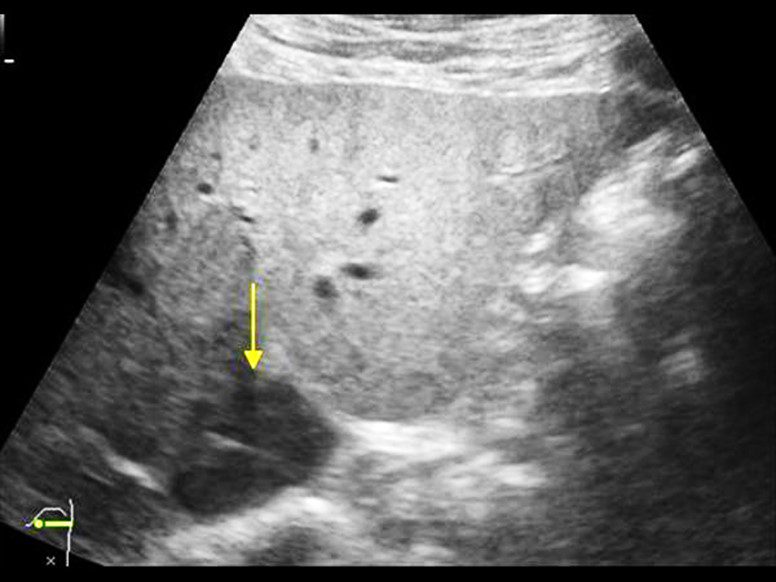
Grey Scale Usg Image Shows A Hypoechoic Nodule Arrow In The Left Lobe
Grey Scale Usg Image Shows A Hypoechoic Nodule Arrow In The Left Lobe
729×601
A Subcapsular Inhomogeneous Hypoechoic Focal Liver Lesion Of 35 ×
A Subcapsular Inhomogeneous Hypoechoic Focal Liver Lesion Of 35 ×
850×587

Ultrasonography Shows A Well Demarcated Partly Exophytic Hypoechoic
Ultrasonography Shows A Well Demarcated Partly Exophytic Hypoechoic
702×697

Mixed Hypoechoic Type Hemangioma Transverse Us Image Of The Liver
Mixed Hypoechoic Type Hemangioma Transverse Us Image Of The Liver
714×513
Ultrasonographic View Of An Abnormal Right Liver Lobe In A 3 Year Old
Ultrasonographic View Of An Abnormal Right Liver Lobe In A 3 Year Old
850×585
Hepatocellular Carcinoma Oval Hypoechoic Lesion 3 Cm In Diameter In
Hepatocellular Carcinoma Oval Hypoechoic Lesion 3 Cm In Diameter In
850×268

Focal Hypoechoic Lesion Snn In A 34 Year Old Man With Fatty Liver A
Focal Hypoechoic Lesion Snn In A 34 Year Old Man With Fatty Liver A
850×913
