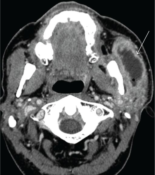
Left Facial Abscess A Axial Contrast Enhanced Ct So Open I

Find inspiration for Left Facial Abscess A Axial Contrast Enhanced Ct So Open I with our image finder website, Left Facial Abscess A Axial Contrast Enhanced Ct So Open I is one of the most popular images and photo galleries in Axial Contrast Enhanced Ct Scan Shows Bilateral Facial Soft Tissue Gallery, Left Facial Abscess A Axial Contrast Enhanced Ct So Open I Picture are available in collection of high-quality images and discover endless ideas for your living spaces, You will be able to watch high quality photo galleries Left Facial Abscess A Axial Contrast Enhanced Ct So Open I.
aiartphotoz.com is free images/photos finder and fully automatic search engine, No Images files are hosted on our server, All links and images displayed on our site are automatically indexed by our crawlers, We only help to make it easier for visitors to find a free wallpaper, background Photos, Design Collection, Home Decor and Interior Design photos in some search engines. aiartphotoz.com is not responsible for third party website content. If this picture is your intelectual property (copyright infringement) or child pornography / immature images, please send email to aiophotoz[at]gmail.com for abuse. We will follow up your report/abuse within 24 hours.
Related Images of Left Facial Abscess A Axial Contrast Enhanced Ct So Open I
Axial Contrast Enhanced Ct Scan Shows Bilateral Facial Soft Tissue
Axial Contrast Enhanced Ct Scan Shows Bilateral Facial Soft Tissue
850×863
Axial Contrast Enhanced Ct Scan Shows Bilateral Facial Soft Tissue
Axial Contrast Enhanced Ct Scan Shows Bilateral Facial Soft Tissue
1024×768
A Axial Contrast Enhanced Ct Image Shows Bilateral Herniation Of The
A Axial Contrast Enhanced Ct Image Shows Bilateral Herniation Of The
850×250
Axial Contrast Enhanced Ct Scan Images With Soft Tissue A And Bone
Axial Contrast Enhanced Ct Scan Images With Soft Tissue A And Bone
640×640
Pre Treatment Axial Contrast Enhanced Ct Scan Images A And B Show An
Pre Treatment Axial Contrast Enhanced Ct Scan Images A And B Show An
640×640
Axial Contrast Enhanced Ct Soft Tissue Window Of The Paranasal Showed
Axial Contrast Enhanced Ct Soft Tissue Window Of The Paranasal Showed
641×491
Left Facial Abscess A Axial Contrast Enhanced Ct So Open I
Left Facial Abscess A Axial Contrast Enhanced Ct So Open I
512×578
Axial Contrast Enhanced Ct Images Showing A Well Demarcated
Axial Contrast Enhanced Ct Images Showing A Well Demarcated
600×481
Contrast Enhanced Ct Scans Showing A Huge Soft Tissue Mass With
Contrast Enhanced Ct Scans Showing A Huge Soft Tissue Mass With
850×748
Axial Contrast Enhanced Ct Soft Tissue Window A Of The Head Shows
Axial Contrast Enhanced Ct Soft Tissue Window A Of The Head Shows
767×442
Axial And Coronal Sinus Contrast Enhanced Ct Scans Showing A
Axial And Coronal Sinus Contrast Enhanced Ct Scans Showing A
850×439
Axial Contrast Enhanced Ct Scan Shows Posterior Bulging Of The
Axial Contrast Enhanced Ct Scan Shows Posterior Bulging Of The
850×819
Showing A Contrast Enhanced Axial Ct Of The Neck Highlighted Area
Showing A Contrast Enhanced Axial Ct Of The Neck Highlighted Area
689×511
Contrast Enhanced Ct Scan In Axial Views Of Neck A 36 × 48 Mm Soft
Contrast Enhanced Ct Scan In Axial Views Of Neck A 36 × 48 Mm Soft
600×693
A Axial Contrast Enhanced Ct Scan Of A 2 Year Old Patient Showing A
A Axial Contrast Enhanced Ct Scan Of A 2 Year Old Patient Showing A
850×436
Orocutaneous Fistula A B Axial Contrast Enhanced Ct Of The Oral
Orocutaneous Fistula A B Axial Contrast Enhanced Ct Of The Oral
640×640
Contrast Enhanced Axial Ct Scan Shows Bilateral Minimal Pleural
Contrast Enhanced Axial Ct Scan Shows Bilateral Minimal Pleural
731×521
A Axial Contrast Enhanced Ct Scan Soft Tissue Window Showing A
A Axial Contrast Enhanced Ct Scan Soft Tissue Window Showing A
600×710
Axial Contrast Enhanced Ct Scan Performed At The Same Level As In 1
Axial Contrast Enhanced Ct Scan Performed At The Same Level As In 1
553×450
Unenhanced Axial Ct And Contrast Enhanced Ct Scans A Unenhanced
Unenhanced Axial Ct And Contrast Enhanced Ct Scans A Unenhanced
567×567
Axial Contrast Enhanced Ct Found Multiloculated Low Attenuation Masses
Axial Contrast Enhanced Ct Found Multiloculated Low Attenuation Masses
850×739
Axial Contrast Enhanced Ct Scan At The Level Of The Floor Of The
Axial Contrast Enhanced Ct Scan At The Level Of The Floor Of The
850×706
A Axial Contrast Enhanced Ct Image With Mediastinal Window Settings
A Axial Contrast Enhanced Ct Image With Mediastinal Window Settings
600×750
A Coronal Ct Scan Shows An Irregular Shape Contrast Enhanced Right
A Coronal Ct Scan Shows An Irregular Shape Contrast Enhanced Right
850×438
B Contrast Enhanced Ct Scan Axial View Showing Soft Tissue Mass In The
B Contrast Enhanced Ct Scan Axial View Showing Soft Tissue Mass In The
622×752
A Series Of Contrast Enhanced Axial Ct Scans Shows On The Right Side A
A Series Of Contrast Enhanced Axial Ct Scans Shows On The Right Side A
640×640
Axial Section Of Contrast Enhanced Ct Scan Showing Hetrogenously
Axial Section Of Contrast Enhanced Ct Scan Showing Hetrogenously
685×527
Axial Contrast Enhanced Ct Scan Demonstrated A Solid Download
Axial Contrast Enhanced Ct Scan Demonstrated A Solid Download
640×640
Axial Contrast Enhanced Ct Scan At The Level Of True Vocal Cords
Axial Contrast Enhanced Ct Scan At The Level Of True Vocal Cords
600×571
Axial Contrast Enhanced Ct Image A Demonstrates An Irregular Soft
Axial Contrast Enhanced Ct Image A Demonstrates An Irregular Soft
850×583
Panels A C Contrast Enhanced Ct Scan Of Soft Tissues Of The Neck
Panels A C Contrast Enhanced Ct Scan Of Soft Tissues Of The Neck
640×640
A B Axial Contrast Enhanced Ct Scans Show A Heterogeneous
A B Axial Contrast Enhanced Ct Scans Show A Heterogeneous
640×640
Ct Scan Features A Axial Contrast Enhanced Section Showing Diffuse
Ct Scan Features A Axial Contrast Enhanced Section Showing Diffuse
788×259
