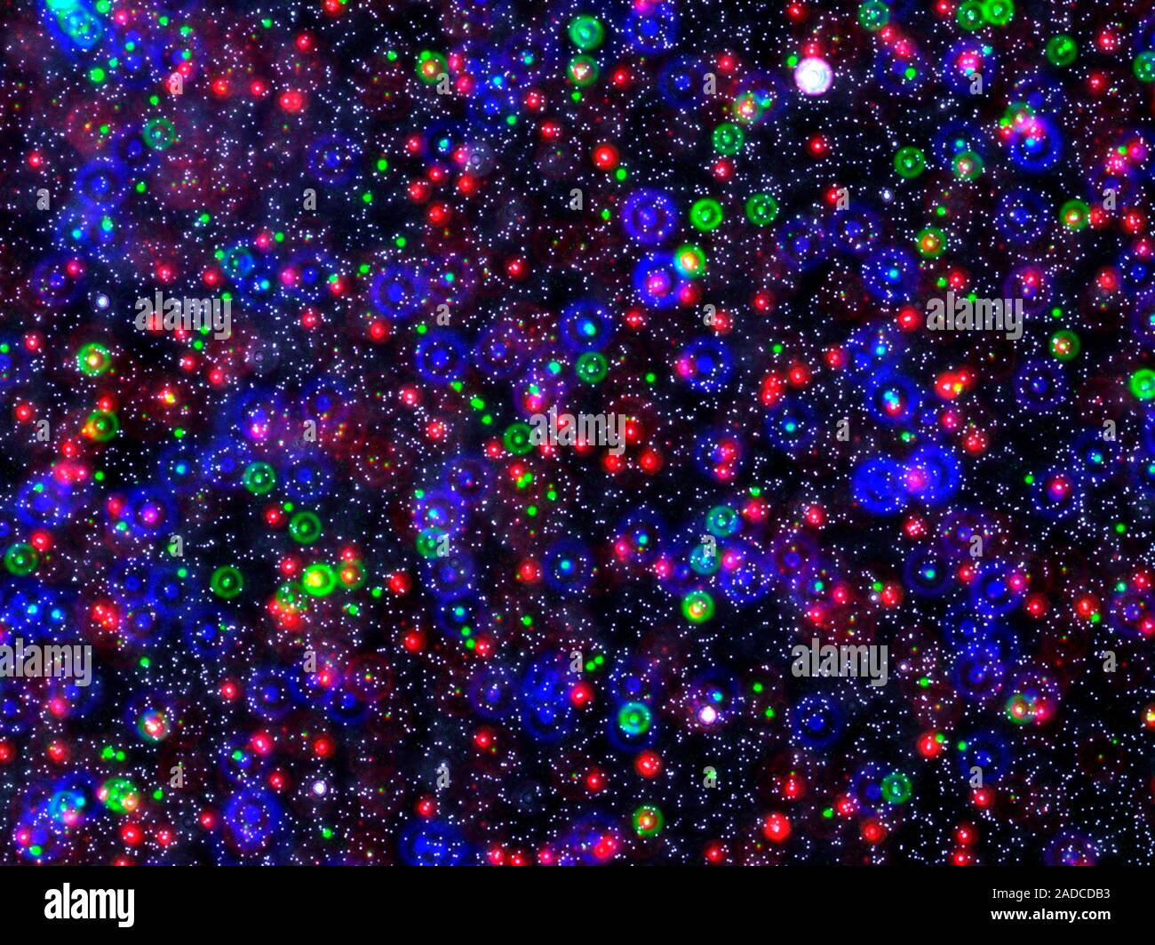
Microsphere Calibration Beads Fluorescent Light Micrograph Of Multi

Find inspiration for Microsphere Calibration Beads Fluorescent Light Micrograph Of Multi with our image finder website, Microsphere Calibration Beads Fluorescent Light Micrograph Of Multi is one of the most popular images and photo galleries in Microsphere Calibration Beads Fluorescent Light Micrograph Of Multi Gallery, Microsphere Calibration Beads Fluorescent Light Micrograph Of Multi Picture are available in collection of high-quality images and discover endless ideas for your living spaces, You will be able to watch high quality photo galleries Microsphere Calibration Beads Fluorescent Light Micrograph Of Multi.
aiartphotoz.com is free images/photos finder and fully automatic search engine, No Images files are hosted on our server, All links and images displayed on our site are automatically indexed by our crawlers, We only help to make it easier for visitors to find a free wallpaper, background Photos, Design Collection, Home Decor and Interior Design photos in some search engines. aiartphotoz.com is not responsible for third party website content. If this picture is your intelectual property (copyright infringement) or child pornography / immature images, please send email to aiophotoz[at]gmail.com for abuse. We will follow up your report/abuse within 24 hours.
Related Images of Microsphere Calibration Beads Fluorescent Light Micrograph Of Multi
Microsphere Calibration Beads Fluorescent Light Micrograph Of Multi
Microsphere Calibration Beads Fluorescent Light Micrograph Of Multi
1300×1063
Microsphere Calibration Beads Fluorescent Light Micrograph Stock
Microsphere Calibration Beads Fluorescent Light Micrograph Stock
800×599
Microsphere Beads Fluorescent Light Micrograph Stock Image C036
Microsphere Beads Fluorescent Light Micrograph Stock Image C036
634×800
Microsphere Beads Fluorescent Light Micrograph Beads Such As These
Microsphere Beads Fluorescent Light Micrograph Beads Such As These
1030×1390
Experimental Results Of The Multimodal System Of Microsphere Beads
Experimental Results Of The Multimodal System Of Microsphere Beads
850×880
Fluorescent Bead Calibration Of The Relative Position Of The Two
Fluorescent Bead Calibration Of The Relative Position Of The Two
850×849
4 Checking Z Positioning Calibration Using Fluorescent Beads A 102
4 Checking Z Positioning Calibration Using Fluorescent Beads A 102
850×843
14 Micrograph Of Fluorescent Beads Imaged Through A 60x Microscope
14 Micrograph Of Fluorescent Beads Imaged Through A 60x Microscope
850×700
Calibration By Fluorescent Beads And Schematic Diagram Of Sample Stage
Calibration By Fluorescent Beads And Schematic Diagram Of Sample Stage
850×739
Morphology Of The Fluorescent Polymer Microspheres Pamba Rhb A The
Morphology Of The Fluorescent Polymer Microspheres Pamba Rhb A The
850×809
Multiple Microsphere Nanoscopes Fluorescent Pictures Showing Multiple
Multiple Microsphere Nanoscopes Fluorescent Pictures Showing Multiple
640×640
Microsphere Control Beads Day 1 Red And Day 7 Green Control Images
Microsphere Control Beads Day 1 Red And Day 7 Green Control Images
458×1132
Fluorescent Microspheres Microparticles And Spherical Powders
Fluorescent Microspheres Microparticles And Spherical Powders
1061×805
Linearized Detection Of Microparticles By Using Calibration Sized Beads
Linearized Detection Of Microparticles By Using Calibration Sized Beads
641×903
Recording Of Fluorescent Beads A In A Light Sheet Microscope As
Recording Of Fluorescent Beads A In A Light Sheet Microscope As
850×950
Fluorescent Microsphere Based Assay Each Bead Contains Two Dyes At
Fluorescent Microsphere Based Assay Each Bead Contains Two Dyes At
550×407
Fluorescent Light Micrograph Of Dna Security Beads Stock Image T980
Fluorescent Light Micrograph Of Dna Security Beads Stock Image T980
800×605
Image Showing Fluorescent Microspheres In Two Axially Distinct Layers
Image Showing Fluorescent Microspheres In Two Axially Distinct Layers
665×1565
Confocal Micrograph Depicting Phagocytosis Of Fluorescent Beads By
Confocal Micrograph Depicting Phagocytosis Of Fluorescent Beads By
537×1085
A A Fluorescence Micrograph Following Bead Dispensing Into Two
A A Fluorescence Micrograph Following Bead Dispensing Into Two
574×856
This Fluorescent Micrograph Shows The Assembly Of 300 Nm Beads On An
This Fluorescent Micrograph Shows The Assembly Of 300 Nm Beads On An
850×608
Depiction Of Encoding Beads With Multiple Fluorescent Dyes To Produce
Depiction Of Encoding Beads With Multiple Fluorescent Dyes To Produce
850×1105
Aggregates Of 43 µm Fluorescent Microsphere Settled At The Bottom Of
Aggregates Of 43 µm Fluorescent Microsphere Settled At The Bottom Of
640×640
Calibration Scans Of Fluorescent Beads A Fluorescence Intensity
Calibration Scans Of Fluorescent Beads A Fluorescence Intensity
640×640
Transmission Electron Micrograph Of Fluorescent Microsphere Fms
Transmission Electron Micrograph Of Fluorescent Microsphere Fms
500×422
Figure3 A Schematic Illustration Of The Signal Recording For The
Figure3 A Schematic Illustration Of The Signal Recording For The
850×593
Integrated Spim Illumination For Fluorescence Imaging A Schematic Of
Integrated Spim Illumination For Fluorescence Imaging A Schematic Of
850×663
This Fluorescent Micrograph Shows The Assembly Of 300 Nm Beads On An
This Fluorescent Micrograph Shows The Assembly Of 300 Nm Beads On An
640×640
Precision Microspheres Beads Microparticles Powder Particle Sizes
Precision Microspheres Beads Microparticles Powder Particle Sizes
640×480
Microsphere Based Super Resolution Endoscopy Imaging In Fluorescence
Microsphere Based Super Resolution Endoscopy Imaging In Fluorescence
585×565
Bead Calibration Of Dls A 41 Mixture Of 102 Nm And 404 Nm Polystyrene
Bead Calibration Of Dls A 41 Mixture Of 102 Nm And 404 Nm Polystyrene
708×558
Light Microscopy Of Beads Size And Appearance Of Different Size Of
Light Microscopy Of Beads Size And Appearance Of Different Size Of
850×317
Illustration Of An Example Of A Bead Or Microsphere Array Used In Bba
Illustration Of An Example Of A Bead Or Microsphere Array Used In Bba
850×481
Image Scanning Microscopy Of Fluorescent Beads A Side By Side
Image Scanning Microscopy Of Fluorescent Beads A Side By Side
850×399
