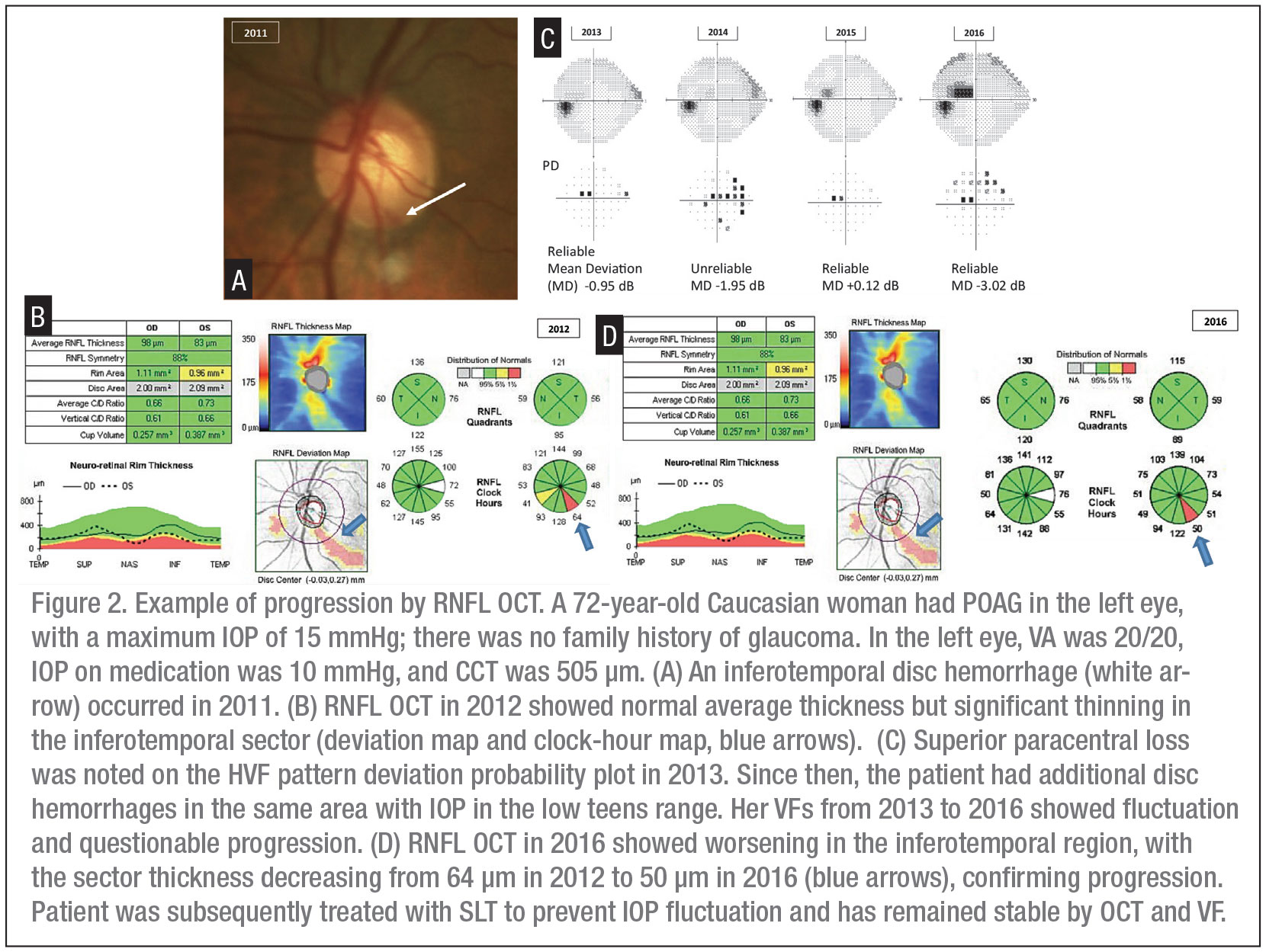
Monitoring Glaucoma Progression With Oct

Find inspiration for Monitoring Glaucoma Progression With Oct with our image finder website, Monitoring Glaucoma Progression With Oct is one of the most popular images and photo galleries in Oct Disc With Glaucoma Gallery, Monitoring Glaucoma Progression With Oct Picture are available in collection of high-quality images and discover endless ideas for your living spaces, You will be able to watch high quality photo galleries Monitoring Glaucoma Progression With Oct.
aiartphotoz.com is free images/photos finder and fully automatic search engine, No Images files are hosted on our server, All links and images displayed on our site are automatically indexed by our crawlers, We only help to make it easier for visitors to find a free wallpaper, background Photos, Design Collection, Home Decor and Interior Design photos in some search engines. aiartphotoz.com is not responsible for third party website content. If this picture is your intelectual property (copyright infringement) or child pornography / immature images, please send email to aiophotoz[at]gmail.com for abuse. We will follow up your report/abuse within 24 hours.
Related Images of Monitoring Glaucoma Progression With Oct
Optical Coherence Tomography Oct Applecross Eye Clinic
Optical Coherence Tomography Oct Applecross Eye Clinic
940×1021
Lesson Maximizing Oct In The Diagnosis And Management Of Glaucoma
Lesson Maximizing Oct In The Diagnosis And Management Of Glaucoma
1200×1364
Atlas Entry Megalopapillae And Severe Glaucoma
Atlas Entry Megalopapillae And Severe Glaucoma
1366×1684
Optic Disc Margin Anatomic Features In Myopic Eyes With Glaucoma With
Optic Disc Margin Anatomic Features In Myopic Eyes With Glaucoma With
2067×1530
Optic Disc Characteristics In Patients With Glaucoma And Combined
Optic Disc Characteristics In Patients With Glaucoma And Combined
1922×1178
Heidelberg Engineering Receives Fda Clearance To Market Spectralis® Oct
Heidelberg Engineering Receives Fda Clearance To Market Spectralis® Oct
1000×964
Acs Eye Specialists Oct Optical Coherence Tomography Used For
Acs Eye Specialists Oct Optical Coherence Tomography Used For
657×335
Lesson Maximizing Oct In The Diagnosis And Management Of Glaucoma
Lesson Maximizing Oct In The Diagnosis And Management Of Glaucoma
1200×719
Automated Glaucoma Detection Using Retinal Layers Segmentation And
Automated Glaucoma Detection Using Retinal Layers Segmentation And
1800×999
Monitoring Glaucoma Progression With Oct 48 Off
Monitoring Glaucoma Progression With Oct 48 Off
1280×720
Series Of Optic Disc Photographs Optical Coherence Tomography Oct
Series Of Optic Disc Photographs Optical Coherence Tomography Oct
850×481
Evaluating The Optic Nerve For Glaucomatous Damage With Oct Glaucoma
Evaluating The Optic Nerve For Glaucomatous Damage With Oct Glaucoma
721×513
Optic Disc Pit Oct Image American Academy Of Ophthalmology
Optic Disc Pit Oct Image American Academy Of Ophthalmology
1000×1000
Sd Oct Findings In A Case Of Glaucoma Suspect She Had A Large Disc
Sd Oct Findings In A Case Of Glaucoma Suspect She Had A Large Disc
600×478
Oct Oct A Useful In Identifying Va Decline In Glaucoma
Oct Oct A Useful In Identifying Va Decline In Glaucoma
596×399
Lesson Understanding Onh Dynamics In Glaucoma And Beyond
Lesson Understanding Onh Dynamics In Glaucoma And Beyond
1410×1110
Lesson Maximizing Oct In The Diagnosis And Management Of Glaucoma
Lesson Maximizing Oct In The Diagnosis And Management Of Glaucoma
1200×808
Hypoperfusion On Oct A Correlates With Vf Progression In Glaucoma
Hypoperfusion On Oct A Correlates With Vf Progression In Glaucoma
1200×701
Spectral Domain Optical Coherence Tomography For Glaucoma Diagnosis
Spectral Domain Optical Coherence Tomography For Glaucoma Diagnosis
600×378
What Is Glaucoma Pietermaritzburg Eye Hospital
What Is Glaucoma Pietermaritzburg Eye Hospital
1024×594
Normal Optic Disc And Glaucomatous Optic Nerve Heads New Glaucoma
Normal Optic Disc And Glaucomatous Optic Nerve Heads New Glaucoma
2283×1063
