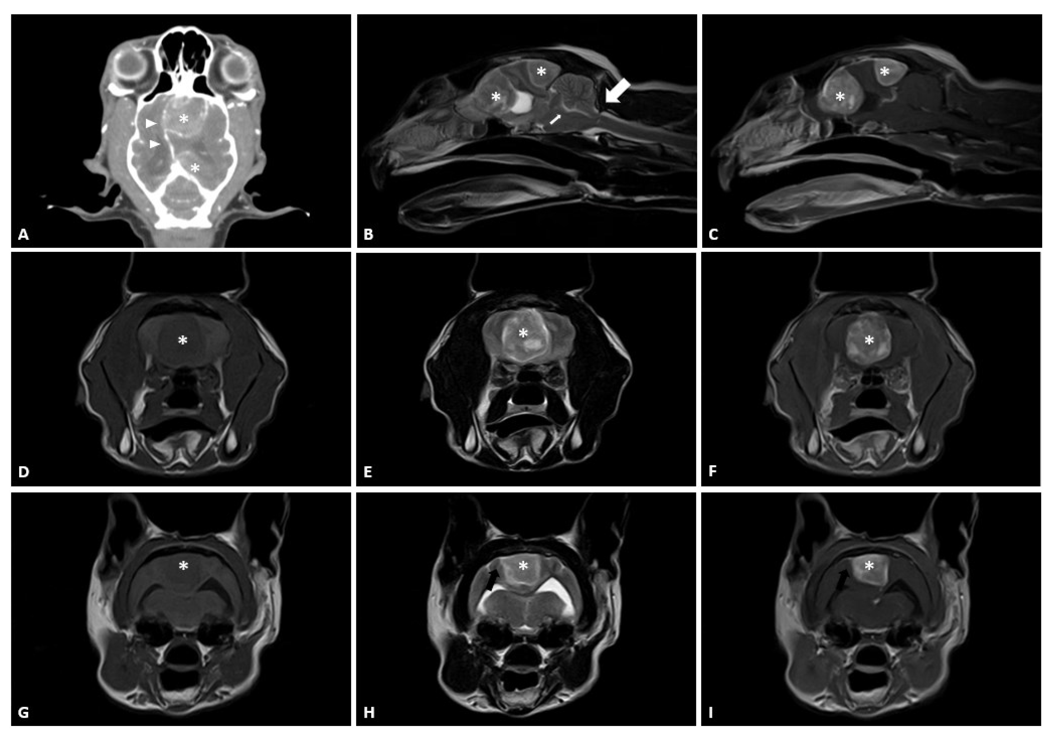
Multiple Meningioma Resection By Bilateral Extended Rostrotentorial

Find inspiration for Multiple Meningioma Resection By Bilateral Extended Rostrotentorial with our image finder website, Multiple Meningioma Resection By Bilateral Extended Rostrotentorial is one of the most popular images and photo galleries in Multiple Meningioma Resection By Bilateral Extended Rostrotentorial Gallery, Multiple Meningioma Resection By Bilateral Extended Rostrotentorial Picture are available in collection of high-quality images and discover endless ideas for your living spaces, You will be able to watch high quality photo galleries Multiple Meningioma Resection By Bilateral Extended Rostrotentorial.
aiartphotoz.com is free images/photos finder and fully automatic search engine, No Images files are hosted on our server, All links and images displayed on our site are automatically indexed by our crawlers, We only help to make it easier for visitors to find a free wallpaper, background Photos, Design Collection, Home Decor and Interior Design photos in some search engines. aiartphotoz.com is not responsible for third party website content. If this picture is your intelectual property (copyright infringement) or child pornography / immature images, please send email to aiophotoz[at]gmail.com for abuse. We will follow up your report/abuse within 24 hours.
Related Images of Multiple Meningioma Resection By Bilateral Extended Rostrotentorial
Pdf Multiple Meningioma Resection By Bilateral Extended
Pdf Multiple Meningioma Resection By Bilateral Extended
850×1202
Multiple Meningioma Resection By Bilateral Extended Rostrotentorial
Multiple Meningioma Resection By Bilateral Extended Rostrotentorial
3354×2347
Multiple Meningioma Resection By Bilateral Extended Rostrotentorial
Multiple Meningioma Resection By Bilateral Extended Rostrotentorial
2980×1327
Figure 3 From The Extended Supracerebellar Transtentorial Approach For
Figure 3 From The Extended Supracerebellar Transtentorial Approach For
482×520
Magnetic Resonance Imaging Of Multiple Meningiomas Arising Within The
Magnetic Resonance Imaging Of Multiple Meningiomas Arising Within The
850×322
Illustration Of A Combined Approach And Using Imri Case 3 A
Illustration Of A Combined Approach And Using Imri Case 3 A
850×907
Figure 2 From Combined Simultaneous Transcranial And Endoscopic
Figure 2 From Combined Simultaneous Transcranial And Endoscopic
674×470
Figure 2 From The Extended Supracerebellar Transtentorial Approach For
Figure 2 From The Extended Supracerebellar Transtentorial Approach For
1076×1128
Figure 9 From The Extended Supracerebellar Transtentorial Approach For
Figure 9 From The Extended Supracerebellar Transtentorial Approach For
346×522
Surgical Treatment Of Rostrotentorial Meningioma Complicated By
Surgical Treatment Of Rostrotentorial Meningioma Complicated By
1230×554
Figure 1 From Successful Resection Of Bilateral Parafalcine Meningioma
Figure 1 From Successful Resection Of Bilateral Parafalcine Meningioma
1002×706
Figure 1 From Bilateral Supplementary Motor Area Syndrome Causing
Figure 1 From Bilateral Supplementary Motor Area Syndrome Causing
674×1466
Mri Images Of Meningioma Before And After Resection Axial Postcontrast
Mri Images Of Meningioma Before And After Resection Axial Postcontrast
850×647
Changes Of The Pre And Post Operative Meningioma Before A And
Changes Of The Pre And Post Operative Meningioma Before A And
640×640
Right Frontotemporal Craniotomy For Resection Of Tuberculum Sella
Right Frontotemporal Craniotomy For Resection Of Tuberculum Sella
2357×1158
Figure 1 From Combined Simultaneous Transcranial And Endoscopic
Figure 1 From Combined Simultaneous Transcranial And Endoscopic
676×512
Figure 3 From Bilateral Supplementary Motor Area Syndrome Causing
Figure 3 From Bilateral Supplementary Motor Area Syndrome Causing
648×1258
Figure 1 From Endoscopic Endonasal Resection Via A Transsphenoidal And
Figure 1 From Endoscopic Endonasal Resection Via A Transsphenoidal And
1228×1422
Xilloc Medical Bv One Stage Meningioma Resection Patient Specific
Xilloc Medical Bv One Stage Meningioma Resection Patient Specific
1920×1200
Parasagittalparafalcine Meningioma Resection Principles Operative
Parasagittalparafalcine Meningioma Resection Principles Operative
640×360
Preoperatively Designed Three Dimensional 3d Guide And Models For
Preoperatively Designed Three Dimensional 3d Guide And Models For
531×531
53f With A Convexity Meningioma Mri A Before And B After Complete
53f With A Convexity Meningioma Mri A Before And B After Complete
850×1029
Figure 3 From Microsurgical Excision Of Multiple Clear Cell Meningiomas
Figure 3 From Microsurgical Excision Of Multiple Clear Cell Meningiomas
1378×670
Cancers Free Full Text Surgical And Functional Outcome After
Cancers Free Full Text Surgical And Functional Outcome After
3488×3963
Figure 4 From Bilateral Supplementary Motor Area Syndrome Causing
Figure 4 From Bilateral Supplementary Motor Area Syndrome Causing
614×1272
Craniotomy And Complete Resection Of Recurrent Parafalcine Meningioma
Craniotomy And Complete Resection Of Recurrent Parafalcine Meningioma
936×486
Figure 1 From Cerebellar And Multiple Spinal Hemangioblastomas And
Figure 1 From Cerebellar And Multiple Spinal Hemangioblastomas And
1314×692
98 Pterional Craniotomy And Extradural Anterior Clinoidectomy For
98 Pterional Craniotomy And Extradural Anterior Clinoidectomy For
3840×2160
Right Frontotemporal Craniotomy For Resection Of Tuberculum Sella
Right Frontotemporal Craniotomy For Resection Of Tuberculum Sella
963×1083
Frontiers Resection Of Olfactory Groove Meningiomas Through
Frontiers Resection Of Olfactory Groove Meningiomas Through
1084×933
Resection Of A Petroclival Meningioma Through The Extended Retrosigmoid
Resection Of A Petroclival Meningioma Through The Extended Retrosigmoid
850×422
Right Frontal Meningioma Pre And Postembolization And Resection
Right Frontal Meningioma Pre And Postembolization And Resection
818×608
Pre And Postoperative Images Of A Patient With A Tuberculum Sellae
Pre And Postoperative Images Of A Patient With A Tuberculum Sellae
1500×668
Anterior Transpetrosal Approach For Resection Of Petroclival Meningioma
Anterior Transpetrosal Approach For Resection Of Petroclival Meningioma
415×569
Caring For Meningiomas A Multi Disciplinary Approach
Caring For Meningiomas A Multi Disciplinary Approach
