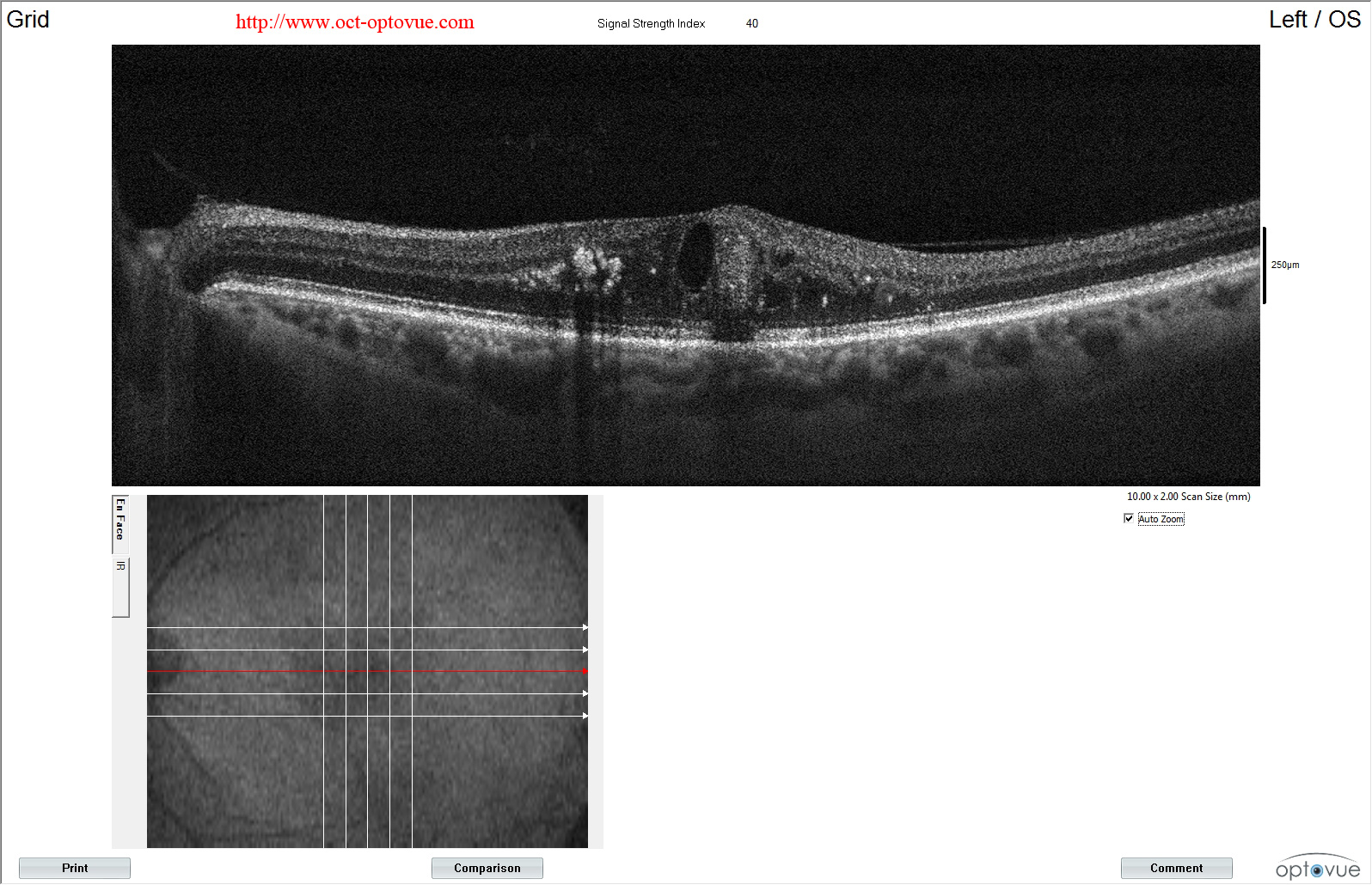
Oct Angiography And Vascular Diseases

Find inspiration for Oct Angiography And Vascular Diseases with our image finder website, Oct Angiography And Vascular Diseases is one of the most popular images and photo galleries in Detecting Retinal Lesions With Oct Dr Jerome Sherman Youtube Gallery, Oct Angiography And Vascular Diseases Picture are available in collection of high-quality images and discover endless ideas for your living spaces, You will be able to watch high quality photo galleries Oct Angiography And Vascular Diseases.
aiartphotoz.com is free images/photos finder and fully automatic search engine, No Images files are hosted on our server, All links and images displayed on our site are automatically indexed by our crawlers, We only help to make it easier for visitors to find a free wallpaper, background Photos, Design Collection, Home Decor and Interior Design photos in some search engines. aiartphotoz.com is not responsible for third party website content. If this picture is your intelectual property (copyright infringement) or child pornography / immature images, please send email to aiophotoz[at]gmail.com for abuse. We will follow up your report/abuse within 24 hours.
Related Images of Oct Angiography And Vascular Diseases
Detecting Retinal Lesions With Oct Dr Jerome Sherman Youtube
Detecting Retinal Lesions With Oct Dr Jerome Sherman Youtube
572×384
How To Read An Oct Image With Dr Jerome Sherman Youtube
How To Read An Oct Image With Dr Jerome Sherman Youtube
1009×799
Oct Scan Retinal Imaging At Eye Solutions The Complete Eye Hospital
Oct Scan Retinal Imaging At Eye Solutions The Complete Eye Hospital
519×340
Oct And Fundus Imaging An Update With Dr Jerome Sherman Youtube
Oct And Fundus Imaging An Update With Dr Jerome Sherman Youtube
512×496
Understanding Autofluorescence Imaging With Dr Jerome Sherman
Understanding Autofluorescence Imaging With Dr Jerome Sherman
1395×922
Detecting Retina Damage From Oct Retinal Images Towards Data Science
Detecting Retina Damage From Oct Retinal Images Towards Data Science
3414×1937
A Complete List Of Ocular Diseases With Optical Coherence Tomography Oct
A Complete List Of Ocular Diseases With Optical Coherence Tomography Oct
790×212
Detecting Retina Damage From Oct Retinal Images Towards Data Science
Detecting Retina Damage From Oct Retinal Images Towards Data Science
3413×1920
4 Tips For Assessing The Macular Oct Scan Artofit
4 Tips For Assessing The Macular Oct Scan Artofit
384×501
The Utility Of En Face Oct For Detecting Neovascularization In Dr
The Utility Of En Face Oct For Detecting Neovascularization In Dr
1200×1606
Practical Utility Of Widefield Oct Angiography To Detect Retinal
Practical Utility Of Widefield Oct Angiography To Detect Retinal
850×952
Detecting Retina Damage From Oct Retinal Images Towards Data Science
Detecting Retina Damage From Oct Retinal Images Towards Data Science
1093×588
Practical Utility Of Widefield Oct Angiography To Detect Retinal
Practical Utility Of Widefield Oct Angiography To Detect Retinal
640×640
The Retinal Nerve Fiber Layer In Clinical Practice Sherman Jerome
The Retinal Nerve Fiber Layer In Clinical Practice Sherman Jerome
771×638
Oct Scans Of Retinal Lesions With Altered Melanin Location A Bone
Oct Scans Of Retinal Lesions With Altered Melanin Location A Bone
1200×954
Dots Spots And Other White Retinal Lesions Page 56 Of 61 Retina
Dots Spots And Other White Retinal Lesions Page 56 Of 61 Retina
1146×1280
Associated Retinal Lesions Sd Oct B Scans Passing Through The Avls Of
Associated Retinal Lesions Sd Oct B Scans Passing Through The Avls Of
640×640
Practical Utility Of Widefield Oct Angiography To Detect Retinal
Practical Utility Of Widefield Oct Angiography To Detect Retinal
498×568
Suspicious Retinal Lesion With A Distinctive Appearance Indian
Suspicious Retinal Lesion With A Distinctive Appearance Indian
1596×1030
Application Of Mydriasis And Eye Steering In Ultrawide Field Imaging
Application Of Mydriasis And Eye Steering In Ultrawide Field Imaging
702×315
Wide Field Swept Source Oct Angiography 23 × 20 Mm For Detecting
Wide Field Swept Source Oct Angiography 23 × 20 Mm For Detecting
736×958
Capability Of The Oct To Detect And Capture The Nature Of Lesions
Capability Of The Oct To Detect And Capture The Nature Of Lesions
1061×677
Oct Scans For The Same Patient With Chronic Retinal Detachment
Oct Scans For The Same Patient With Chronic Retinal Detachment
758×390
Lesson Peripheral Retinal Imaging And Disease Assessment
Lesson Peripheral Retinal Imaging And Disease Assessment
861×1024
Retinal Ischemic Perivascular Lesions Ripls Are Characterized By
Retinal Ischemic Perivascular Lesions Ripls Are Characterized By
850×884
Detecting Retina Damage From Oct Retinal Images Towards Data Science
Detecting Retina Damage From Oct Retinal Images Towards Data Science
1085×573
Retinoschisisretinal Detachment Page 12 Of 42 Retina Revealed
Retinoschisisretinal Detachment Page 12 Of 42 Retina Revealed
Pdf Detecting Red Lesions From Retinal Fundus Images Using Unique
Pdf Detecting Red Lesions From Retinal Fundus Images Using Unique
Oct And Retinal Digital Imaging Feltham Eyecare Centre
Oct And Retinal Digital Imaging Feltham Eyecare Centre
