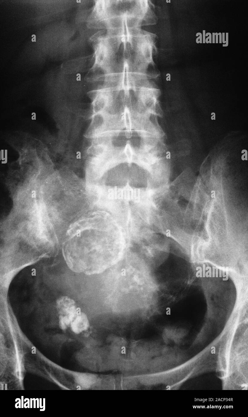
Pelvic X Ray Front View Showing Calcified Cysts In A Womans Ovary
Find inspiration for Pelvic X Ray Front View Showing Calcified Cysts In A Womans Ovary with our image finder website, Pelvic X Ray Front View Showing Calcified Cysts In A Womans Ovary is one of the most popular images and photo galleries in X Ray Of The Pelvis Showing Calcifications In Bilateral Common Iliac Gallery, Pelvic X Ray Front View Showing Calcified Cysts In A Womans Ovary Picture are available in collection of high-quality images and discover endless ideas for your living spaces, You will be able to watch high quality photo galleries Pelvic X Ray Front View Showing Calcified Cysts In A Womans Ovary.
aiartphotoz.com is free images/photos finder and fully automatic search engine, No Images files are hosted on our server, All links and images displayed on our site are automatically indexed by our crawlers, We only help to make it easier for visitors to find a free wallpaper, background Photos, Design Collection, Home Decor and Interior Design photos in some search engines. aiartphotoz.com is not responsible for third party website content. If this picture is your intelectual property (copyright infringement) or child pornography / immature images, please send email to aiophotoz[at]gmail.com for abuse. We will follow up your report/abuse within 24 hours.
Related Images of Pelvic X Ray Front View Showing Calcified Cysts In A Womans Ovary

X Ray Of The Pelvis Showing Calcifications In Bilateral Common Iliac
X Ray Of The Pelvis Showing Calcifications In Bilateral Common Iliac
665×526

Abnormal Calcifications In A Pelvic Radiograph The Bmj
Abnormal Calcifications In A Pelvic Radiograph The Bmj
1645×2000

Small Calcifications In The Pelvis On X Ray Radiology In Plain English
Small Calcifications In The Pelvis On X Ray Radiology In Plain English
773×1030
Sadaf Iftikhar Mbbs Fcps Mayo Clinic Proceedings Ppt Download
Sadaf Iftikhar Mbbs Fcps Mayo Clinic Proceedings Ppt Download
1024×768

Diagnostic Approach To Benign And Malignant Calcifications In The
Diagnostic Approach To Benign And Malignant Calcifications In The
1303×1239

Anteroposterior Ap Radiograph Of The Pelvis Shows Calcification Of
Anteroposterior Ap Radiograph Of The Pelvis Shows Calcification Of
612×524
Dystrophic Calcifications Secondary To Intramuscular Injections
Dystrophic Calcifications Secondary To Intramuscular Injections
3028×2472


Ap Pelvic X Ray Demonstrating Bilateral Sacroiliitis And Hip
Ap Pelvic X Ray Demonstrating Bilateral Sacroiliitis And Hip
715×494

Diagnostic Approach To Benign And Malignant Calcifications In The
Diagnostic Approach To Benign And Malignant Calcifications In The
1599×1327

Abdominal And Pelvic X Ray Shows Multiple Calcified Soft Tissue
Abdominal And Pelvic X Ray Shows Multiple Calcified Soft Tissue
850×1031

Small Calcifications In The Pelvis On X Ray Radiology In Plain English
Small Calcifications In The Pelvis On X Ray Radiology In Plain English
845×321

Xray Of The Pelvis With Bilateral Hips In Anteroposterior View Showing
Xray Of The Pelvis With Bilateral Hips In Anteroposterior View Showing
713×505
Pediatric Pelvis Trauma Radiographic Evaluation Pediatrics Orthobullets
Pediatric Pelvis Trauma Radiographic Evaluation Pediatrics Orthobullets
685×534

Conventional X Ray Pelvis Anteroposterior Projection Revealed Right
Conventional X Ray Pelvis Anteroposterior Projection Revealed Right
850×697

Pelvic X Ray Front View Showing Calcified Cysts In A Womans Ovary
Pelvic X Ray Front View Showing Calcified Cysts In A Womans Ovary
829×1390

Plain Abdominal X Rays Showing A Calcified Mass In The Pelvis
Plain Abdominal X Rays Showing A Calcified Mass In The Pelvis
410×549

The Hip Bone Ilium Ischium Pubis Teachmeanatomy
The Hip Bone Ilium Ischium Pubis Teachmeanatomy
1107×906

Vascular Calcifications And Aneurysms Radiology Key
Vascular Calcifications And Aneurysms Radiology Key
500×245


X Ray Of Normal Pelvis Female Eccles Health Sciences Library J
X Ray Of Normal Pelvis Female Eccles Health Sciences Library J
2140×1848
Anterior Posterior Pelvic X Rays Exhibiting Bilateral Injection
Anterior Posterior Pelvic X Rays Exhibiting Bilateral Injection
850×265
X Ray Pelvis Of The Patient Showing Bilateral Sacroilitis Download
X Ray Pelvis Of The Patient Showing Bilateral Sacroilitis Download
753×516


X Ray Pelvis Showing Small Square Iliac Crest With Spikes Of Bone At
X Ray Pelvis Showing Small Square Iliac Crest With Spikes Of Bone At
555×555

Abdominal X Ray Abnormal Calcification Vascular Calcification
Abdominal X Ray Abnormal Calcification Vascular Calcification
900×565

Diagnostic Approach To Benign And Malignant Calcifications In The
Diagnostic Approach To Benign And Malignant Calcifications In The
455×500

Abdominal X Ray Of The Patient Showing Clustered Calcifications In
Abdominal X Ray Of The Patient Showing Clustered Calcifications In
693×958

Diagnostic Approach To Benign And Malignant Calcifications In The
Diagnostic Approach To Benign And Malignant Calcifications In The
500×478

Pelvic X Ray Stock Image P1160713 Science Photo Library
Pelvic X Ray Stock Image P1160713 Science Photo Library
800×646
Plain Abdominal X Rays Showing A Calcified Mass In The Pelvis
Plain Abdominal X Rays Showing A Calcified Mass In The Pelvis
539×539

Hip X Ray Interpretation Osce Guide Geeky Medics
Hip X Ray Interpretation Osce Guide Geeky Medics
1536×864
Calcification Of The Aorta And Common Iliac Arteries Nejm
Calcification Of The Aorta And Common Iliac Arteries Nejm
1200×452

Diagnostic Approach To Benign And Malignant Calcifications In The
Diagnostic Approach To Benign And Malignant Calcifications In The
1550×1183
