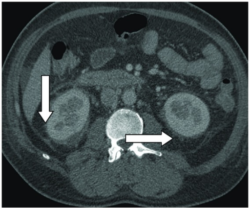
Perinephric Fat Stranding Ultrasound

Find inspiration for Perinephric Fat Stranding Ultrasound with our image finder website, Perinephric Fat Stranding Ultrasound is one of the most popular images and photo galleries in Perinephric Fat Stranding Ultrasound Gallery, Perinephric Fat Stranding Ultrasound Picture are available in collection of high-quality images and discover endless ideas for your living spaces, You will be able to watch high quality photo galleries Perinephric Fat Stranding Ultrasound.
aiartphotoz.com is free images/photos finder and fully automatic search engine, No Images files are hosted on our server, All links and images displayed on our site are automatically indexed by our crawlers, We only help to make it easier for visitors to find a free wallpaper, background Photos, Design Collection, Home Decor and Interior Design photos in some search engines. aiartphotoz.com is not responsible for third party website content. If this picture is your intelectual property (copyright infringement) or child pornography / immature images, please send email to aiophotoz[at]gmail.com for abuse. We will follow up your report/abuse within 24 hours.
Related Images of Perinephric Fat Stranding Ultrasound
Figure 1 From Para And Perirenal Fat Thickness Is An Independent
Figure 1 From Para And Perirenal Fat Thickness Is An Independent
896×568
Pdf Non Invasive Prediction Of Carotid Artery Atherosclerosis By
Pdf Non Invasive Prediction Of Carotid Artery Atherosclerosis By
505×505
Echocardiographic Perirenal Fat Thickness The Perirenal Fat Located
Echocardiographic Perirenal Fat Thickness The Perirenal Fat Located
640×640
Noncontrast Ct Kub Showing Enlarged Right Kidney With Perinephric Fat
Noncontrast Ct Kub Showing Enlarged Right Kidney With Perinephric Fat
640×640
Ultrasound Showing Distended Appendix With Surrounding Fat Stranding
Ultrasound Showing Distended Appendix With Surrounding Fat Stranding
850×506
Computed Tomography Axial View Perinephric Fat Stranding Around Both
Computed Tomography Axial View Perinephric Fat Stranding Around Both
850×675
Frontiers Perirenal Fat Volume Is Positively Associated With Serum
Frontiers Perirenal Fat Volume Is Positively Associated With Serum
4094×1385
Abdominal Ultrasound Of 58 Years Old Female Patient Showing A The
Abdominal Ultrasound Of 58 Years Old Female Patient Showing A The
850×619
Ultrasound Showing A Large Perinephric Haematoma Measuring About 96 ×
Ultrasound Showing A Large Perinephric Haematoma Measuring About 96 ×
850×622
Method Of Measuring Of Posterior Perinephric Fat Thickness At The Level
Method Of Measuring Of Posterior Perinephric Fat Thickness At The Level
640×640
Ultrasonography Of Peritoneal And Retroperitoneal Spaces And Abdominal
Ultrasonography Of Peritoneal And Retroperitoneal Spaces And Abdominal
895×600
Ultrasound Examination Of The Left And Right Common Carotid Arteries Of
Ultrasound Examination Of The Left And Right Common Carotid Arteries Of
640×640
Differential Diagnosis Of Perinephric Masses On Ct And Mri Ajr
Differential Diagnosis Of Perinephric Masses On Ct And Mri Ajr
1280×907
Measurement Of Perinephric Fat At The Level Of The Renal Vein
Measurement Of Perinephric Fat At The Level Of The Renal Vein
671×503
Noncontrast Ct Kub Showing Enlarged Right Kidney With Perinephric Fat
Noncontrast Ct Kub Showing Enlarged Right Kidney With Perinephric Fat
640×640
As Shown By Non Contrast Enhanced Ct Perirenal Fat Stranding
As Shown By Non Contrast Enhanced Ct Perirenal Fat Stranding
373×760
Ultrasound Assessed Perirenal Fat Is Related To Increased Ophthalmic
Ultrasound Assessed Perirenal Fat Is Related To Increased Ophthalmic
785×640
Gray Scale Ultrasound Selected Images Of The Transplanted Kidney And
Gray Scale Ultrasound Selected Images Of The Transplanted Kidney And
850×365
