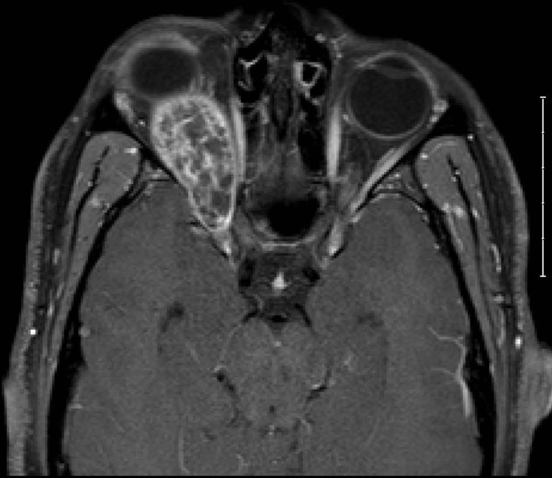
Rare Case Of Orbital Schwannoma With Intralesional Haemorrhage And

Find inspiration for Rare Case Of Orbital Schwannoma With Intralesional Haemorrhage And with our image finder website, Rare Case Of Orbital Schwannoma With Intralesional Haemorrhage And is one of the most popular images and photo galleries in Figure 1 From Pediatric Dumbbell Shaped Orbital Schwannoma With Gallery, Rare Case Of Orbital Schwannoma With Intralesional Haemorrhage And Picture are available in collection of high-quality images and discover endless ideas for your living spaces, You will be able to watch high quality photo galleries Rare Case Of Orbital Schwannoma With Intralesional Haemorrhage And.
aiartphotoz.com is free images/photos finder and fully automatic search engine, No Images files are hosted on our server, All links and images displayed on our site are automatically indexed by our crawlers, We only help to make it easier for visitors to find a free wallpaper, background Photos, Design Collection, Home Decor and Interior Design photos in some search engines. aiartphotoz.com is not responsible for third party website content. If this picture is your intelectual property (copyright infringement) or child pornography / immature images, please send email to aiophotoz[at]gmail.com for abuse. We will follow up your report/abuse within 24 hours.
Related Images of Rare Case Of Orbital Schwannoma With Intralesional Haemorrhage And
Figure 1 From Pediatric Dumbbell Shaped Orbital Schwannoma With
Figure 1 From Pediatric Dumbbell Shaped Orbital Schwannoma With
1340×716
Frontiers Pediatric Dumbbell Shaped Orbital Schwannoma With Extension
Frontiers Pediatric Dumbbell Shaped Orbital Schwannoma With Extension
1248×1256
Figure 1 From Micro Neurosurgical Excision Of Dumbbell Shaped Very
Figure 1 From Micro Neurosurgical Excision Of Dumbbell Shaped Very
526×604
Pdf Pediatric Dumbbell Shaped Orbital Schwannoma With Extension To
Pdf Pediatric Dumbbell Shaped Orbital Schwannoma With Extension To
850×1203
Frontiers Pediatric Dumbbell Shaped Orbital Schwannoma With Extension
Frontiers Pediatric Dumbbell Shaped Orbital Schwannoma With Extension
1248×836
Frontiers Pediatric Dumbbell Shaped Orbital Schwannoma With Extension
Frontiers Pediatric Dumbbell Shaped Orbital Schwannoma With Extension
1248×941
Figure 1 From Schwannoma Of The Orbit Semantic Scholar
Figure 1 From Schwannoma Of The Orbit Semantic Scholar
500×427
Figure 1 From Giant Dumbbell Shaped Intra And Extracranial Nerve
Figure 1 From Giant Dumbbell Shaped Intra And Extracranial Nerve
1132×254
T1 Weighted Mri Sequences With Contrast Enhancement Show A Right
T1 Weighted Mri Sequences With Contrast Enhancement Show A Right
850×267
A T1 With Contrast Schwannoma Shows Enhancement And B T2wi Shows A
A T1 With Contrast Schwannoma Shows Enhancement And B T2wi Shows A
850×513
Figure 1 From Orbital Schwannoma Extending To The Lateral Wall Of The
Figure 1 From Orbital Schwannoma Extending To The Lateral Wall Of The
1032×330
Figure 1 From Surgical Considerations For Maximal Safe Excision Of
Figure 1 From Surgical Considerations For Maximal Safe Excision Of
1028×566
Hybrid Endoscopic Microscopic Surgery For Dumbbell Shaped Trigeminal
Hybrid Endoscopic Microscopic Surgery For Dumbbell Shaped Trigeminal
1294×1010
Figure 1 From Micro Neurosurgical Excision Of Dumbbell Shaped Very
Figure 1 From Micro Neurosurgical Excision Of Dumbbell Shaped Very
526×626
Giant Spinal Thoracic Dumbbell Schwannoma In Pediatric
Giant Spinal Thoracic Dumbbell Schwannoma In Pediatric
1800×592
Imaging Findings Of Pediatric Orbital Masses And Tumor Mimics
Imaging Findings Of Pediatric Orbital Masses And Tumor Mimics
500×250
Figure 1 From Surgical Resection Of Thoracic Dumbbell Shaped Schwannoma
Figure 1 From Surgical Resection Of Thoracic Dumbbell Shaped Schwannoma
1024×462
Rare Case Of Orbital Schwannoma With Intralesional Haemorrhage And
Rare Case Of Orbital Schwannoma With Intralesional Haemorrhage And
1729×1800
Figure 1 From A Sporadic Pediatric Case Of A Spinal Dumbbell Shaped
Figure 1 From A Sporadic Pediatric Case Of A Spinal Dumbbell Shaped
944×646
Axial Mr Images Demonstrating A Medium Sized Dumbbell Shaped
Axial Mr Images Demonstrating A Medium Sized Dumbbell Shaped
850×324
Rare Case Of Orbital Schwannoma With Intralesional Haemorrhage And
Rare Case Of Orbital Schwannoma With Intralesional Haemorrhage And
1800×1559
Figure 1 From Acute Neurological Aggravation Caused By Intratumoral
Figure 1 From Acute Neurological Aggravation Caused By Intratumoral
946×1518
Figure 2 From Dumbbell Shaped Primitive Neuroectodermal Tumor Mimicking
Figure 2 From Dumbbell Shaped Primitive Neuroectodermal Tumor Mimicking
1260×1632
Figure 1 From Pediatric Eighth Cranial Nerve Schwannoma Without
Figure 1 From Pediatric Eighth Cranial Nerve Schwannoma Without
1428×880
Figure 1 From Giant Recurrent Dumbbell Shaped Hypoglossal Schwannoma In
Figure 1 From Giant Recurrent Dumbbell Shaped Hypoglossal Schwannoma In
1164×396
Figure 1 From Micro Neurosurgical Excision Of Dumbbell Shaped Very
Figure 1 From Micro Neurosurgical Excision Of Dumbbell Shaped Very
526×398
Giant Orbital Schwannoma With Fluidfluid Levels British Journal Of
Giant Orbital Schwannoma With Fluidfluid Levels British Journal Of
825×1800
Figure 1 From A Dumbbell Shaped Meningioma Mimicking A Schwannoma In
Figure 1 From A Dumbbell Shaped Meningioma Mimicking A Schwannoma In
632×354
Figure 1 From Une Localisation Rare Dun Schwannome Orbitaire A Rare
Figure 1 From Une Localisation Rare Dun Schwannome Orbitaire A Rare
1290×746
Preoperative Mri Revealed A Right C3c4 Dumbbell Shaped Foraminal
Preoperative Mri Revealed A Right C3c4 Dumbbell Shaped Foraminal
850×470
Giant Dumbbell Shaped Schwannoma But Not Transforaminal
Giant Dumbbell Shaped Schwannoma But Not Transforaminal
882×1280
Vestibular Schwannomas Diagnosis And Surgical Treatment Intechopen
Vestibular Schwannomas Diagnosis And Surgical Treatment Intechopen
591×361
Microsurgical Resection Of A Right Dumbbell Shaped Jugular Foramen
Microsurgical Resection Of A Right Dumbbell Shaped Jugular Foramen
685×576
