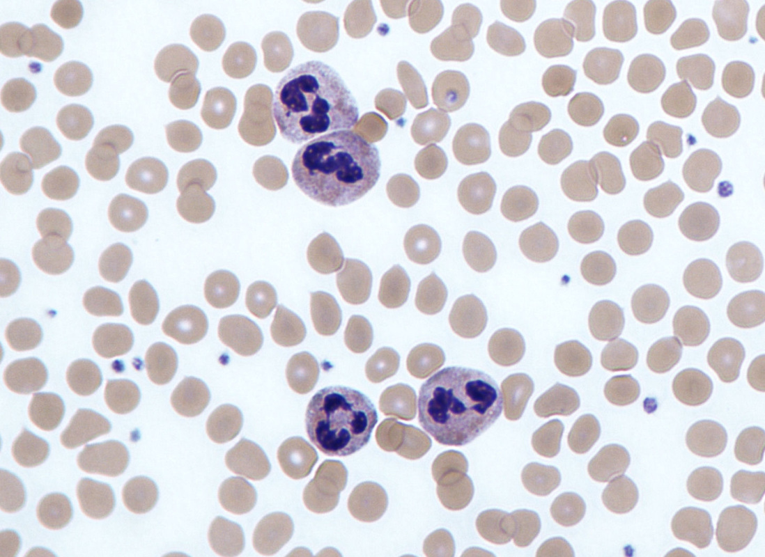
Rbcs Wbcs Secs In Gram Stains Microbiology Learning The Whyology

Find inspiration for Rbcs Wbcs Secs In Gram Stains Microbiology Learning The Whyology with our image finder website, Rbcs Wbcs Secs In Gram Stains Microbiology Learning The Whyology is one of the most popular images and photo galleries in Neutrophils Gram Stain Gallery, Rbcs Wbcs Secs In Gram Stains Microbiology Learning The Whyology Picture are available in collection of high-quality images and discover endless ideas for your living spaces, You will be able to watch high quality photo galleries Rbcs Wbcs Secs In Gram Stains Microbiology Learning The Whyology.
aiartphotoz.com is free images/photos finder and fully automatic search engine, No Images files are hosted on our server, All links and images displayed on our site are automatically indexed by our crawlers, We only help to make it easier for visitors to find a free wallpaper, background Photos, Design Collection, Home Decor and Interior Design photos in some search engines. aiartphotoz.com is not responsible for third party website content. If this picture is your intelectual property (copyright infringement) or child pornography / immature images, please send email to aiophotoz[at]gmail.com for abuse. We will follow up your report/abuse within 24 hours.
Related Images of Rbcs Wbcs Secs In Gram Stains Microbiology Learning The Whyology
Gram Stain Tutorial Microbiology Learning The Whyology Of
Gram Stain Tutorial Microbiology Learning The Whyology Of
1079×800
Sputum Gram Stain Shown Many Neutrophils Stock Photo 1050410753
Sputum Gram Stain Shown Many Neutrophils Stock Photo 1050410753
1500×1450
Gram Negative Diplococci Neutrophils Stained By Stock Photo 646804351
Gram Negative Diplococci Neutrophils Stained By Stock Photo 646804351
1479×1600
Cerebrospinal Fluid Csf Gram Stain Reveals Numerous Neutrophils The
Cerebrospinal Fluid Csf Gram Stain Reveals Numerous Neutrophils The
753×509
Sputum Gram Stain Shown Many Neutrophils库存照片1050410750 Shutterstock
Sputum Gram Stain Shown Many Neutrophils库存照片1050410750 Shutterstock
1500×1464
Gram Stain Of Swab Exudate Answer 4 Arrows Point Towards Neutrophils
Gram Stain Of Swab Exudate Answer 4 Arrows Point Towards Neutrophils
850×655
Sputum Gram Stain Shown Many Neutrophils Stock Photo 1050410756
Sputum Gram Stain Shown Many Neutrophils Stock Photo 1050410756
1500×1455
Rbcs Wbcs Secs In Gram Stains Microbiology Learning The Whyology
Rbcs Wbcs Secs In Gram Stains Microbiology Learning The Whyology
1098×800
Neutrophil Cell White Blood Cell Peripheral Stock Photo 411399973
Neutrophil Cell White Blood Cell Peripheral Stock Photo 411399973
1500×1225
This Sputum Gram Stain Shown Numerous Stock Photo 1050408680 Shutterstock
This Sputum Gram Stain Shown Numerous Stock Photo 1050408680 Shutterstock
1500×1317
This Sputum Gram Stain Shown Numerous Stock Photo 1050408683 Shutterstock
This Sputum Gram Stain Shown Numerous Stock Photo 1050408683 Shutterstock
1500×1410
Gram Stain Smear Of Ear Secretion Showing Two Polymorphonuclear
Gram Stain Smear Of Ear Secretion Showing Two Polymorphonuclear
648×498
Morphology Of Neutrophils In Hematoxylin And Eosin Stained Sections At
Morphology Of Neutrophils In Hematoxylin And Eosin Stained Sections At
850×608
Gram Stain Image Showing Gram Negative A Xylosoxidans Black Arrows
Gram Stain Image Showing Gram Negative A Xylosoxidans Black Arrows
850×743
Gram Stain Of Wound Specimen Medical Laboratories
Gram Stain Of Wound Specimen Medical Laboratories
828×485
Wright Giemsa Stain Of Csf Demonstrating N Fowleri Organisms And
Wright Giemsa Stain Of Csf Demonstrating N Fowleri Organisms And
850×668
Solved Plate 5 Cerebrospinal Fluid Csf Cytocentrifuge Preparation
Solved Plate 5 Cerebrospinal Fluid Csf Cytocentrifuge Preparation
660×405
Light Micrographs Of Peripheral Blood Cells With Phagocytosis Of Gram
Light Micrographs Of Peripheral Blood Cells With Phagocytosis Of Gram
850×544
Gram Stain Of Sputum Specimen Medical Laboratories
Gram Stain Of Sputum Specimen Medical Laboratories
837×520
Histopathologic Image Of The Tissue Specimen Highlights Thin
Histopathologic Image Of The Tissue Specimen Highlights Thin
520×383
Immunoflogosis Staining With May Grunwald Giemsa Mgg Method Allows
Immunoflogosis Staining With May Grunwald Giemsa Mgg Method Allows
520×668
Gram Staining Of The Sputum Sample A Large Number Of Gram Negative
Gram Staining Of The Sputum Sample A Large Number Of Gram Negative
720×512
