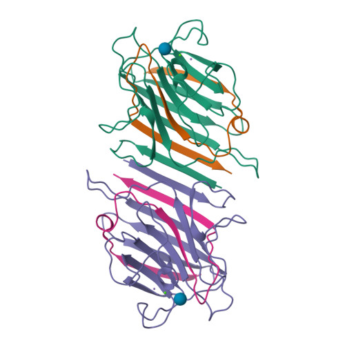
Rcsb Pdb 1lem The Monosaccharide Binding Site Of Lentil Lectin An X

Find inspiration for Rcsb Pdb 1lem The Monosaccharide Binding Site Of Lentil Lectin An X with our image finder website, Rcsb Pdb 1lem The Monosaccharide Binding Site Of Lentil Lectin An X is one of the most popular images and photo galleries in Rcsb Pdb 1lem The Monosaccharide Binding Site Of Lentil Lectin An X Gallery, Rcsb Pdb 1lem The Monosaccharide Binding Site Of Lentil Lectin An X Picture are available in collection of high-quality images and discover endless ideas for your living spaces, You will be able to watch high quality photo galleries Rcsb Pdb 1lem The Monosaccharide Binding Site Of Lentil Lectin An X.
aiartphotoz.com is free images/photos finder and fully automatic search engine, No Images files are hosted on our server, All links and images displayed on our site are automatically indexed by our crawlers, We only help to make it easier for visitors to find a free wallpaper, background Photos, Design Collection, Home Decor and Interior Design photos in some search engines. aiartphotoz.com is not responsible for third party website content. If this picture is your intelectual property (copyright infringement) or child pornography / immature images, please send email to aiophotoz[at]gmail.com for abuse. We will follow up your report/abuse within 24 hours.
Related Images of Rcsb Pdb 1lem The Monosaccharide Binding Site Of Lentil Lectin An X
Rcsb Pdb 1lem The Monosaccharide Binding Site Of Lentil Lectin An X
Rcsb Pdb 1lem The Monosaccharide Binding Site Of Lentil Lectin An X
500×500
Rcsb Pdb 1lem The Monosaccharide Binding Site Of Lentil Lectin An X
Rcsb Pdb 1lem The Monosaccharide Binding Site Of Lentil Lectin An X
500×500
The Monosaccharide And Metal Binding Sites Of Lentil Lectin Pdb Entry
The Monosaccharide And Metal Binding Sites Of Lentil Lectin Pdb Entry
850×593
Rcsb Pdb 1lem The Monosaccharide Binding Site Of Lentil Lectin An X
Rcsb Pdb 1lem The Monosaccharide Binding Site Of Lentil Lectin An X
500×500
Rcsb Pdb 1lob Three Dimensional Structures Of Complexes Of Lathyrus
Rcsb Pdb 1lob Three Dimensional Structures Of Complexes Of Lathyrus
500×500
Rcsb Pdb 1lob Three Dimensional Structures Of Complexes Of Lathyrus
Rcsb Pdb 1lob Three Dimensional Structures Of Complexes Of Lathyrus
500×500
Binding Sites Of Sucrose In Pal And Lentil Lectin A Omplex Of
Binding Sites Of Sucrose In Pal And Lentil Lectin A Omplex Of
850×850
Rcsb Pdb 7xfa Structure Of Human Galectin 3 Crd In Complex With
Rcsb Pdb 7xfa Structure Of Human Galectin 3 Crd In Complex With
2500×2500
A Useful Guide To Lectin Binding Machine Learning Directed Annotation
A Useful Guide To Lectin Binding Machine Learning Directed Annotation
2095×745
Rcsb Pdb 4iab Crystal Structure Of A Putative Monosaccharide Binding
Rcsb Pdb 4iab Crystal Structure Of A Putative Monosaccharide Binding
500×500
Rcsb Pdb 7wmv Structure Of Human Sglt1 Map17 Complex Bound With Lx2761
Rcsb Pdb 7wmv Structure Of Human Sglt1 Map17 Complex Bound With Lx2761
500×500
Rcsb Pdb 8efc Structure Of Lates Calcarifer Dna Polymerase Theta
Rcsb Pdb 8efc Structure Of Lates Calcarifer Dna Polymerase Theta
500×500
Rcsb Pdb 8c87 Double Mutant Al172cll246c Structure Of
Rcsb Pdb 8c87 Double Mutant Al172cll246c Structure Of
2500×2500
Rcsb Pdb 8k2k Crystal Structure Of Group 3 Oligosaccharide
Rcsb Pdb 8k2k Crystal Structure Of Group 3 Oligosaccharide
500×500
Rcsb Pdb 7wbz Crystal Structure Of The Sars Cov 2 Rbd In Complex
Rcsb Pdb 7wbz Crystal Structure Of The Sars Cov 2 Rbd In Complex
500×500
Rcsb Pdb 6lcx Closslinked Alphani Betani Human Hemoglobin A In
Rcsb Pdb 6lcx Closslinked Alphani Betani Human Hemoglobin A In
2500×2500
Rcsb Pdb 4heg Crystal Structure Of Hiv 1 Protease Mutants R8q
Rcsb Pdb 4heg Crystal Structure Of Hiv 1 Protease Mutants R8q
2500×2500
Rcsb Pdb 8c5g Structure Of Human Neuropilin 1 B1b2 Domains In
Rcsb Pdb 8c5g Structure Of Human Neuropilin 1 B1b2 Domains In
500×500
Rcsb Pdb 2f4b Crystal Structure Of The Ligand Binding Domain Of
Rcsb Pdb 2f4b Crystal Structure Of The Ligand Binding Domain Of
500×500
Rcsb Pdb 7vn6 Crystal Structure Of Mbp Fused Bil1bzr1 21 90 In
Rcsb Pdb 7vn6 Crystal Structure Of Mbp Fused Bil1bzr1 21 90 In
500×500
Rcsb Pdb 7mxw Crystal Structure Of Human Exonuclease 1 Exo1 Wt In
Rcsb Pdb 7mxw Crystal Structure Of Human Exonuclease 1 Exo1 Wt In
500×500
Rcsb Pdb 7w7z Crystal Structure Of Human Focal Adhesion Targeting
Rcsb Pdb 7w7z Crystal Structure Of Human Focal Adhesion Targeting
500×500
Rcsb Pdb 7vig Cryo Em Structure Of Gi Coupled Sphingosine 1
Rcsb Pdb 7vig Cryo Em Structure Of Gi Coupled Sphingosine 1
500×500
Rcsb Pdb 7ubi Solution Nmr Structure Of 8 Residue Rosetta Designed
Rcsb Pdb 7ubi Solution Nmr Structure Of 8 Residue Rosetta Designed
500×500
Rcsb Pdb 7we3 Solution Structures Of A Disulfide Rich Peptide That
Rcsb Pdb 7we3 Solution Structures Of A Disulfide Rich Peptide That
500×500
Rcsb Pdb 1ofz Crystal Structure Of Fungal Lectin Six Bladed Beta
Rcsb Pdb 1ofz Crystal Structure Of Fungal Lectin Six Bladed Beta
4485×1824
Rcsb Pdb 7qcu Structure Of The Mucin 2 Cterminal Domains Partially
Rcsb Pdb 7qcu Structure Of The Mucin 2 Cterminal Domains Partially
500×500
Rcsb Pdb 7ph8 Structure Of Insulin Like Growth Factor 1 Receptors
Rcsb Pdb 7ph8 Structure Of Insulin Like Growth Factor 1 Receptors
500×500
Rcsb Pdb 8itu Sars Cov 2 Omicron Ba1 Spike Glycoprotein In Complex
Rcsb Pdb 8itu Sars Cov 2 Omicron Ba1 Spike Glycoprotein In Complex
500×500
Rcsb Pdb 1t5m Structural Transitions As Determinants Of The Action
Rcsb Pdb 1t5m Structural Transitions As Determinants Of The Action
500×500
Rcsb Pdb 4qq8 Crystal Structure Of The Formolase Fls In Space Group
Rcsb Pdb 4qq8 Crystal Structure Of The Formolase Fls In Space Group
2500×2500
Rcsb Pdb 7xfa Structure Of Human Galectin 3 Crd In Complex With
Rcsb Pdb 7xfa Structure Of Human Galectin 3 Crd In Complex With
4860×1835
Rcsb Pdb 1et4 Crystal Structure Of A Vitamin B12 Binding Rna Aptamer
Rcsb Pdb 1et4 Crystal Structure Of A Vitamin B12 Binding Rna Aptamer
500×500
