Sebaceous Cyst Epidermal Cyst And Lipoma Removal In Singapore
Find inspiration for Sebaceous Cyst Epidermal Cyst And Lipoma Removal In Singapore with our image finder website, Sebaceous Cyst Epidermal Cyst And Lipoma Removal In Singapore is one of the most popular images and photo galleries in Epidermoid Cyst Diagram Gallery, Sebaceous Cyst Epidermal Cyst And Lipoma Removal In Singapore Picture are available in collection of high-quality images and discover endless ideas for your living spaces, You will be able to watch high quality photo galleries Sebaceous Cyst Epidermal Cyst And Lipoma Removal In Singapore.
aiartphotoz.com is free images/photos finder and fully automatic search engine, No Images files are hosted on our server, All links and images displayed on our site are automatically indexed by our crawlers, We only help to make it easier for visitors to find a free wallpaper, background Photos, Design Collection, Home Decor and Interior Design photos in some search engines. aiartphotoz.com is not responsible for third party website content. If this picture is your intelectual property (copyright infringement) or child pornography / immature images, please send email to aiophotoz[at]gmail.com for abuse. We will follow up your report/abuse within 24 hours.
Related Images of Sebaceous Cyst Epidermal Cyst And Lipoma Removal In Singapore
Overview Of Epidermoid Cyst European Journal Of Radiology Open
Overview Of Epidermoid Cyst European Journal Of Radiology Open
2917×1794


Epidermoid Cyst Diagram
Epidermoid Cyst Diagram
600×397

Epidermoid Cyst Diagram
Epidermoid Cyst Diagram
850×650
Epidermal Infundibular Cysts Dermatology Advisor
Epidermal Infundibular Cysts Dermatology Advisor
800×600
Epidermoid Cyst Diagram
Epidermoid Cyst Diagram
2600×1199
Epidermoid Cysts In The Hand C J Lincoski D C Bush S J Millon
Epidermoid Cysts In The Hand C J Lincoski D C Bush S J Millon
657×621

Histopathologic Examination An Epidermoid Cyst Is Lined By A Squamous
Histopathologic Examination An Epidermoid Cyst Is Lined By A Squamous
714×476

Histopathological Features Of Epidermoid Cysts Ecs Hematoxylin
Histopathological Features Of Epidermoid Cysts Ecs Hematoxylin
850×955
Histopathology Of Epidermoid Cyst Download Scientific Diagram
Histopathology Of Epidermoid Cyst Download Scientific Diagram
628×480


Epidermoid Cysts A C And D F And Dermoid Cyst G I Case 1 A C
Epidermoid Cysts A C And D F And Dermoid Cyst G I Case 1 A C
850×517
Epidermal Infundibular Cysts Dermatology Advisor
Epidermal Infundibular Cysts Dermatology Advisor
800×600


Tonsillar Epidermoid Cyst On Hpe Black Arrow Dilated Cysts Filled With
Tonsillar Epidermoid Cyst On Hpe Black Arrow Dilated Cysts Filled With
526×526
Overview Of Epidermoid Cyst European Journal Of Radiology Open
Overview Of Epidermoid Cyst European Journal Of Radiology Open
2500×1554

Epidermoid Cyst Histopathological Characteristics A Cystic Type
Epidermoid Cyst Histopathological Characteristics A Cystic Type
477×647

Epidermoid Cysts Treatment In Poughkeepsie Ny Art Of Skin
Epidermoid Cysts Treatment In Poughkeepsie Ny Art Of Skin
1200×1200
A The Appearance Of An Uninfected Epidermoid Cyst B Gross Surgical
A The Appearance Of An Uninfected Epidermoid Cyst B Gross Surgical
850×410

Epidermoid Cyst Vs Sebaceous Cyst What To Expect From A Cyst Removal
Epidermoid Cyst Vs Sebaceous Cyst What To Expect From A Cyst Removal
638×359
Histopathology Determined An Epidermal Cyst Lined By Stratified
Histopathology Determined An Epidermal Cyst Lined By Stratified
687×510

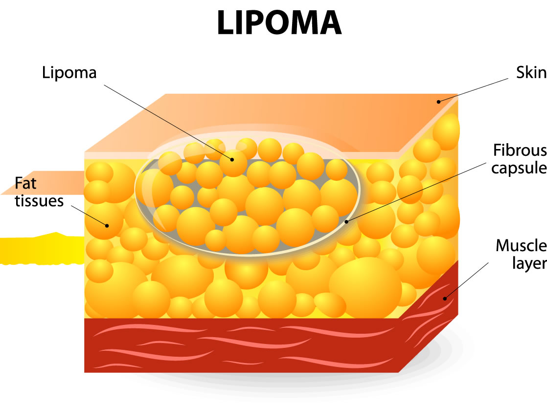
Sebaceous Cyst Epidermal Cyst And Lipoma Removal In Singapore
Sebaceous Cyst Epidermal Cyst And Lipoma Removal In Singapore
1118×835


Characteristic Sonographic Features Of An Epidermoid Cyst Hypoechoic
Characteristic Sonographic Features Of An Epidermoid Cyst Hypoechoic
640×640

Popping A Sebaceous Cyst Can I Burst A Sebaceous Cyst At Home
Popping A Sebaceous Cyst Can I Burst A Sebaceous Cyst At Home
1280×732

Photomicrographs Of Epidermoid Cysts With A Squamous Epithelial Lining
Photomicrographs Of Epidermoid Cysts With A Squamous Epithelial Lining
850×515

Microscopic Image Of A Proliferating Epidermoid Cyst A Type Of
Microscopic Image Of A Proliferating Epidermoid Cyst A Type Of
1000×562
Epidermal Cyst Showing Stratified Squamous Epithelium With Keratin
Epidermal Cyst Showing Stratified Squamous Epithelium With Keratin
812×304
Epidermal Sebaceous Cyst
Epidermal Sebaceous Cyst
750×750

Overview Of Epidermoid Cyst European Journal Of Radiology Open
Overview Of Epidermoid Cyst European Journal Of Radiology Open
2917×1158
