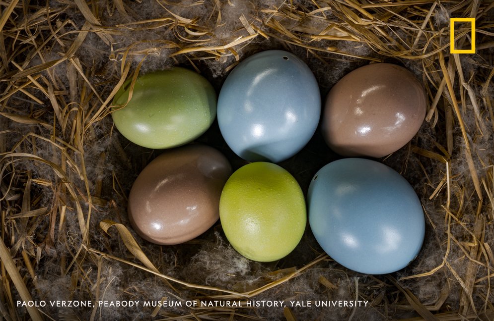
Thread By Natgeo A Microscope Image Of A Quail Embryos Forelimb

Find inspiration for Thread By Natgeo A Microscope Image Of A Quail Embryos Forelimb with our image finder website, Thread By Natgeo A Microscope Image Of A Quail Embryos Forelimb is one of the most popular images and photo galleries in Thread By Natgeo A Microscope Image Of A Quail Embryos Forelimb Gallery, Thread By Natgeo A Microscope Image Of A Quail Embryos Forelimb Picture are available in collection of high-quality images and discover endless ideas for your living spaces, You will be able to watch high quality photo galleries Thread By Natgeo A Microscope Image Of A Quail Embryos Forelimb.
aiartphotoz.com is free images/photos finder and fully automatic search engine, No Images files are hosted on our server, All links and images displayed on our site are automatically indexed by our crawlers, We only help to make it easier for visitors to find a free wallpaper, background Photos, Design Collection, Home Decor and Interior Design photos in some search engines. aiartphotoz.com is not responsible for third party website content. If this picture is your intelectual property (copyright infringement) or child pornography / immature images, please send email to aiophotoz[at]gmail.com for abuse. We will follow up your report/abuse within 24 hours.
Related Images of Thread By Natgeo A Microscope Image Of A Quail Embryos Forelimb
Quail Embryos External Morphology Development Note Quail Embryos With
Quail Embryos External Morphology Development Note Quail Embryos With
640×640
Normal Quail Development The Normal Development Of A Quail Embryo Over
Normal Quail Development The Normal Development Of A Quail Embryo Over
850×666
Thread By Natgeo A Microscope Image Of A Quail Embryos Forelimb
Thread By Natgeo A Microscope Image Of A Quail Embryos Forelimb
995×648
Photomicrographs Of 3 And 4 Day Old Quail Embryos Sectioned During
Photomicrographs Of 3 And 4 Day Old Quail Embryos Sectioned During
600×413
The Top View A And The Side View B Of The 6 Day Quail Embryo
The Top View A And The Side View B Of The 6 Day Quail Embryo
640×640
The Top View A And The Side View B Of The 6 Day Quail Embryo
The Top View A And The Side View B Of The 6 Day Quail Embryo
850×486
A And B Transverse Sections Of Quail Embryos Incubated With The Qh 1
A And B Transverse Sections Of Quail Embryos Incubated With The Qh 1
850×1154
Photomicrographs Of 3 And 4 Day Old Quail Embryos Sectioned During
Photomicrographs Of 3 And 4 Day Old Quail Embryos Sectioned During
600×484
Whole Mounts Of Tenascin Stained Chick Embryos A B And A Quail
Whole Mounts Of Tenascin Stained Chick Embryos A B And A Quail
850×1127
Tunel Wholemounts Of Normal And A− Quail Embryos Brown Cells Are
Tunel Wholemounts Of Normal And A− Quail Embryos Brown Cells Are
751×707
A C Ednrb Expression In Whole Mount Hybridization In Quail Embryos
A C Ednrb Expression In Whole Mount Hybridization In Quail Embryos
850×995
Immunohistochemical Staining Of The Neck Skin Of Quail Embryos At Day 8
Immunohistochemical Staining Of The Neck Skin Of Quail Embryos At Day 8
640×640
Microscopic Image Of A Sagittal Section Of A Control Japanese Quail
Microscopic Image Of A Sagittal Section Of A Control Japanese Quail
640×640
Examples Of Embryos A Hh14 Quail Embryo And B Hh25 Chicken
Examples Of Embryos A Hh14 Quail Embryo And B Hh25 Chicken
539×470
Whole Mounts Of Tenascin Stained Chick Embryos A B And A Quail
Whole Mounts Of Tenascin Stained Chick Embryos A B And A Quail
589×589
Sample Preparation For Imaging Quail Embryos In Vitro A Curved
Sample Preparation For Imaging Quail Embryos In Vitro A Curved
571×571
Photomicrographs Of 3 And 4 Day Old Quail Embryos Sectioned During
Photomicrographs Of 3 And 4 Day Old Quail Embryos Sectioned During
600×447
Photomicrographs Of 6 And 8 Day Old Quail Embryos Sectioned During
Photomicrographs Of 6 And 8 Day Old Quail Embryos Sectioned During
559×450
In Vivo Vascular Network In The Quail Embyro Laser Confocal Microscope
In Vivo Vascular Network In The Quail Embyro Laser Confocal Microscope
850×583
Microscopic Pictures Of 38 H Quail Embryos Ventral View Magnification
Microscopic Pictures Of 38 H Quail Embryos Ventral View Magnification
575×396
Acoustic Images C Scans Of The 11 Day Quail Embryo Head At The Depth
Acoustic Images C Scans Of The 11 Day Quail Embryo Head At The Depth
640×640
Quail Embryo At 10d Incubation 2013 Small World In Motion Competition
Quail Embryo At 10d Incubation 2013 Small World In Motion Competition
640×477
Thread By Natgeo A Microscope Image Of A Quail Embryos Forelimb
Thread By Natgeo A Microscope Image Of A Quail Embryos Forelimb
580×864
Photomicrographs Of 3 Day Old Quail Embryos Sectioned During Genital
Photomicrographs Of 3 Day Old Quail Embryos Sectioned During Genital
600×395
Thread By Natgeo A Microscope Image Of A Quail Embryos Forelimb
Thread By Natgeo A Microscope Image Of A Quail Embryos Forelimb
995×648
Panel A Shows A Quail Embryo Imaged With A Stereomicroscope At 12x
Panel A Shows A Quail Embryo Imaged With A Stereomicroscope At 12x
748×434
Development Of A Quail Embryo Derived By Icsi A Quail Embryo At Handh
Development Of A Quail Embryo Derived By Icsi A Quail Embryo At Handh
729×687
Thread By Natgeo A Microscope Image Of A Quail Embryos Forelimb
Thread By Natgeo A Microscope Image Of A Quail Embryos Forelimb
995×648
Thread By Natgeo A Microscope Image Of A Quail Embryos Forelimb
Thread By Natgeo A Microscope Image Of A Quail Embryos Forelimb
995×648
Thread By Natgeo A Microscope Image Of A Quail Embryos Forelimb
Thread By Natgeo A Microscope Image Of A Quail Embryos Forelimb
995×648
Photomicrographs Of 11 And 14 Day Old Quail Embryos Sectioned During
Photomicrographs Of 11 And 14 Day Old Quail Embryos Sectioned During
532×324
Photomicrographs Of 6 And 8 Day Old Quail Embryos Sectioned During
Photomicrographs Of 6 And 8 Day Old Quail Embryos Sectioned During
600×364
Early Vascularization Of The Cervical Nt In E4 Quail Embryos A C
Early Vascularization Of The Cervical Nt In E4 Quail Embryos A C
727×706
Light Microscopy Of Infected And Uninfected Quail Embryo Cells A
Light Microscopy Of Infected And Uninfected Quail Embryo Cells A
640×640
