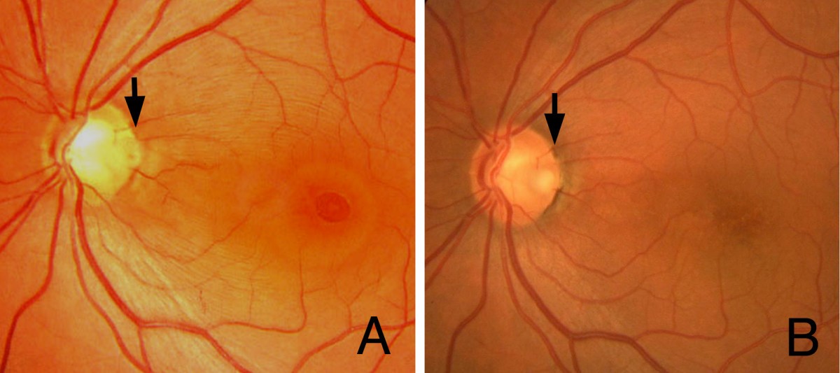
Vitrectomy Combined With Glial Tissue Removal At The Optic Pit In A

Find inspiration for Vitrectomy Combined With Glial Tissue Removal At The Optic Pit In A with our image finder website, Vitrectomy Combined With Glial Tissue Removal At The Optic Pit In A is one of the most popular images and photo galleries in Vitrectomy Combined With Glial Tissue Removal At The Optic Pit In A Gallery, Vitrectomy Combined With Glial Tissue Removal At The Optic Pit In A Picture are available in collection of high-quality images and discover endless ideas for your living spaces, You will be able to watch high quality photo galleries Vitrectomy Combined With Glial Tissue Removal At The Optic Pit In A.
aiartphotoz.com is free images/photos finder and fully automatic search engine, No Images files are hosted on our server, All links and images displayed on our site are automatically indexed by our crawlers, We only help to make it easier for visitors to find a free wallpaper, background Photos, Design Collection, Home Decor and Interior Design photos in some search engines. aiartphotoz.com is not responsible for third party website content. If this picture is your intelectual property (copyright infringement) or child pornography / immature images, please send email to aiophotoz[at]gmail.com for abuse. We will follow up your report/abuse within 24 hours.
Related Images of Vitrectomy Combined With Glial Tissue Removal At The Optic Pit In A
Vitrectomy Combined With Glial Tissue Removal At The Optic Pit In A
Vitrectomy Combined With Glial Tissue Removal At The Optic Pit In A
685×304
Vitrectomy Combined With Glial Tissue Removal At The Optic Pit In A
Vitrectomy Combined With Glial Tissue Removal At The Optic Pit In A
785×351
Pdf Pars Plana Vitrectomy With Air Tamponade For Optic Disc Pit
Pdf Pars Plana Vitrectomy With Air Tamponade For Optic Disc Pit
640×640
Oct Image Seven Years After Vitrectomy Cross Section Of The Temporal
Oct Image Seven Years After Vitrectomy Cross Section Of The Temporal
600×359
Distinguishing Glial Tissue From Optic Disc In Bergmeisters Papilla
Distinguishing Glial Tissue From Optic Disc In Bergmeisters Papilla
1842×1568
Morphologic Changes After Vitrectomy In A Patient With Combined
Morphologic Changes After Vitrectomy In A Patient With Combined
708×534
Glial Tissue Fundus Photographs Of Optic Disc Glial Tissue In Two
Glial Tissue Fundus Photographs Of Optic Disc Glial Tissue In Two
850×604
Figure 3f A Fragment Of Metal Upper Photo Punctures The Eye And
Figure 3f A Fragment Of Metal Upper Photo Punctures The Eye And
500×400
Photographs Of The Right Eye In A 24 Year Old Optic Disc Pit Odp
Photographs Of The Right Eye In A 24 Year Old Optic Disc Pit Odp
571×253
Photographs Of The Right Eye In A 20 Year Old Optic Disc Pit Odp
Photographs Of The Right Eye In A 20 Year Old Optic Disc Pit Odp
574×354
Left Eye Vacant Disc With Coloboma And Fibroglial Tissue Over The Optic
Left Eye Vacant Disc With Coloboma And Fibroglial Tissue Over The Optic
681×440
Real Life Example Of Applicaiton Of Face Down Anterior Vitrectomy A
Real Life Example Of Applicaiton Of Face Down Anterior Vitrectomy A
850×2028
Photographs Of The Right Eye In A 24 Year Old Optic Disc Pit Odp
Photographs Of The Right Eye In A 24 Year Old Optic Disc Pit Odp
571×286
A Observational Management Of Optic Pit Maculopathy Opm Of The Left
A Observational Management Of Optic Pit Maculopathy Opm Of The Left
850×377
Vitrectomy For Diabetic Retinopathy 152 Vitreoretinal Surgery
Vitrectomy For Diabetic Retinopathy 152 Vitreoretinal Surgery
800×740
Figure 1 From Membrane Tissue On The Optic Disc May Cause Macular
Figure 1 From Membrane Tissue On The Optic Disc May Cause Macular
634×1028
Congenital Optic Nerve Pit American Academy Of Ophthalmology
Congenital Optic Nerve Pit American Academy Of Ophthalmology
1201×798
Surgical Procedures Of Staged Lensectomy A C And Vitrectomy D I A
Surgical Procedures Of Staged Lensectomy A C And Vitrectomy D I A
850×427
Challenges In Optic Disc Pit Maculopathy Treatment Retina Today
Challenges In Optic Disc Pit Maculopathy Treatment Retina Today
952×286
Photographs Of The Right Eye In A 20 Year Old Optic Disc Pit Odp
Photographs Of The Right Eye In A 20 Year Old Optic Disc Pit Odp
600×339
Vitrectomy Epiretinal Membrane Removal Youtube
Vitrectomy Epiretinal Membrane Removal Youtube
593×526
Glial Tissue Proliferation On Optic Disc 1 Year Follow Up Download
Glial Tissue Proliferation On Optic Disc 1 Year Follow Up Download
500×334
Full Article Optic Disk Pit Maculopathy Current Management Strategies
Full Article Optic Disk Pit Maculopathy Current Management Strategies
656×1124
Figure 1 From Transconjunctival Sutureless Vitrectomy With Tissue
Figure 1 From Transconjunctival Sutureless Vitrectomy With Tissue
643×438
Accumulation Of Glial Tissue Retina Image Bank
Accumulation Of Glial Tissue Retina Image Bank
862×368
Photographs Of The Left Eye In A 70 Year Old Patient With Maculopathy
Photographs Of The Left Eye In A 70 Year Old Patient With Maculopathy
600×465
Photographs Of The Left Eye In A 70 Year Old Patient With Maculopathy
Photographs Of The Left Eye In A 70 Year Old Patient With Maculopathy
858×642
Congenital And Developmental Anomalies Of The Optic Nerve Ento Key
Congenital And Developmental Anomalies Of The Optic Nerve Ento Key
563×1024
Vitrectomy Without Laser Treatment Or Gas Tamponade For Macular
Vitrectomy Without Laser Treatment Or Gas Tamponade For Macular
367×540
